A 40-year-old man finishes a heavy leg day, weighted squats, leg presses, neck braced back under load. Thirty minutes later: a headache on one side of his skull, vomiting, and he can't swallow right.
Nothing about that list looks neurological until you realize it's not four separate problems. It's one small wedge of brainstem tissue losing its blood supply, and every symptom is a different structure in that wedge announcing exactly where it lives.
Every cranial nerve nucleus has a fixed home. The medulla houses nuclei for cranial nerves VIII, IX, X, XI, and XII. The pons, one level up, houses V, VI, VII, and VIII instead, which is why swallowing trouble points down to the medulla, not up to the pons.
In this case, the exam is a roll call of three of those five. Vestibular signals from cranial nerve VIII misfire, and the room spins. Cranial nerves IX and X share one nucleus, the nucleus ambiguus, so when it fails, both go down together: the swallow breaks and the voice goes hoarse in the same stroke, because it's one nucleus failing, not two.
Those two nuclei sit near the midline. Their silence is not a gap in the exam. It's the clue that tells you exactly where the lesion stops.
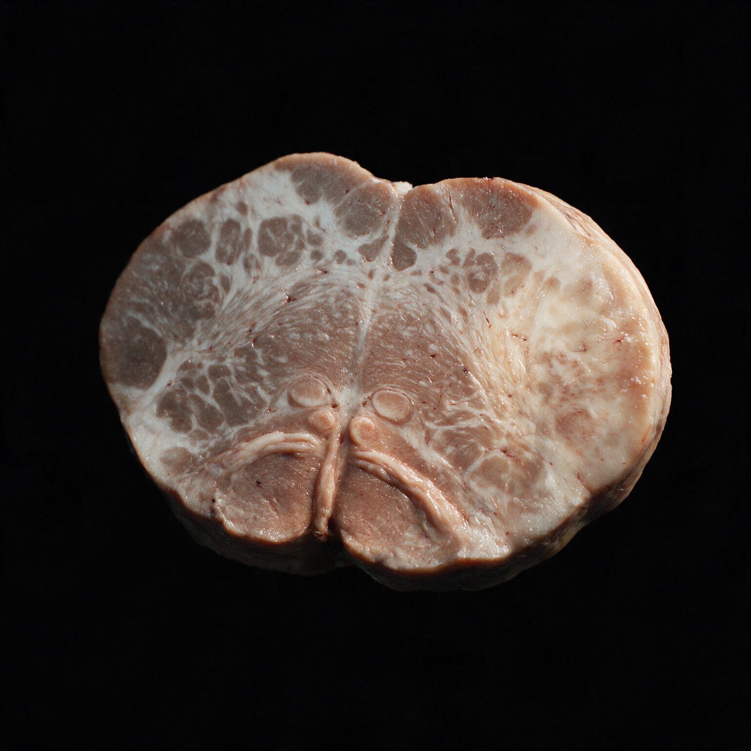
The hypoglossal nucleus and the corticospinal pyramids both sit near the midline of the medulla. Everything else in this case sits laterally: the vestibular nuclei, the nucleus ambiguus, the spinal trigeminal tract carrying facial pain and temperature, the spinothalamic tract carrying body pain and temperature (already crossed lower down, so it reports from the opposite side), the descending sympathetic fibers behind Horner's triad of a drooping eyelid, a small pupil, and dry skin, and the inferior cerebellar peduncle behind the unsteady gait.
This stroke hit the lateral zone and spared the medial zone. That's the entire reason it's named lateral medullary syndrome. Flip it, a medial-zone stroke instead, and you get the mirror image: tongue deviation, weakness on the opposite limb, and none of the vestibular, Horner's, or crossed-sensation findings that define this case.
One detail in this case only makes sense once you see where the nervous system physically crosses its wires: pain and temperature loss on the same side of the face, but the opposite side of the body. That's not two different mechanisms. It's one crossing point, seen from two different tracts that cross at two different levels.
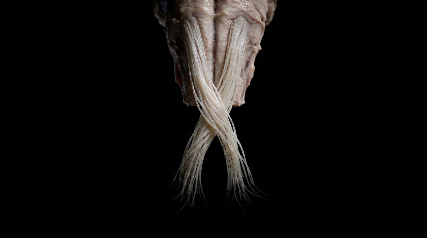
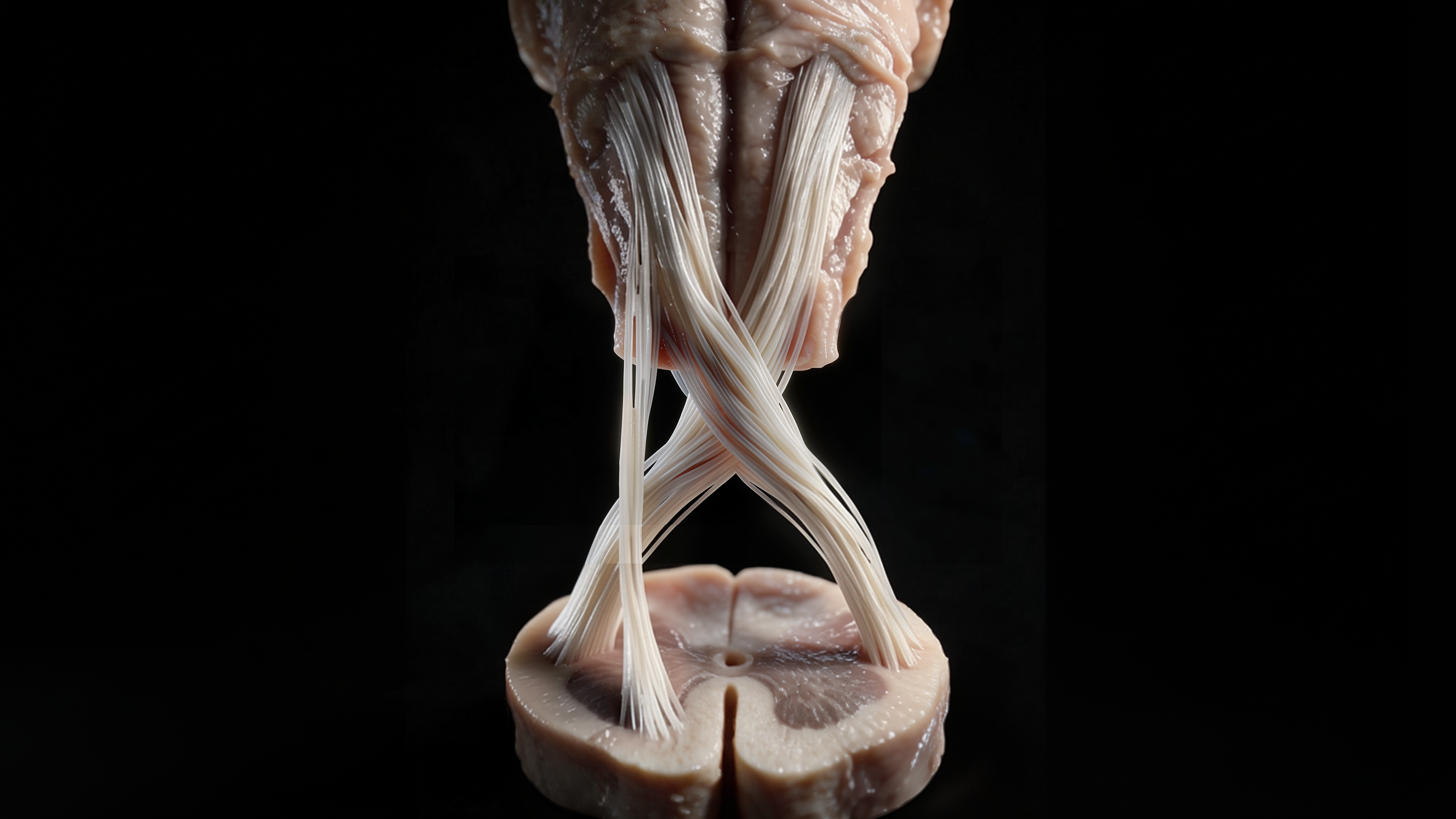
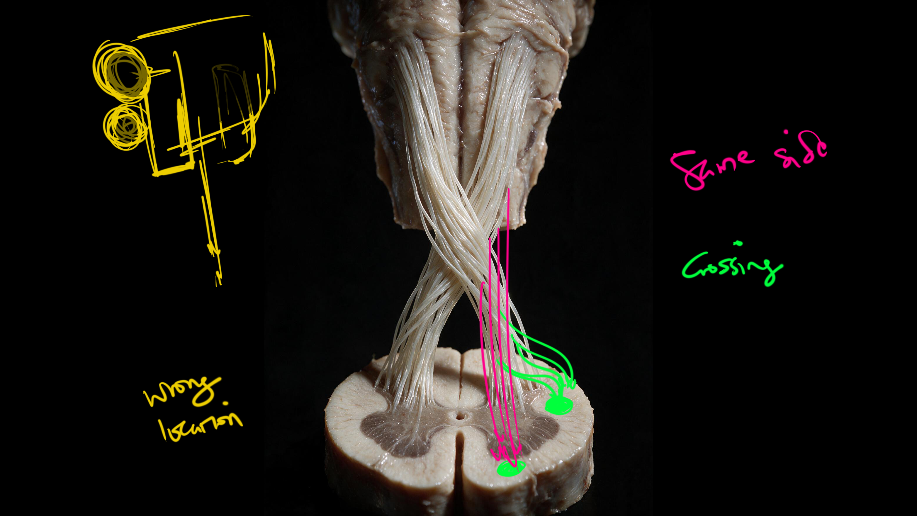
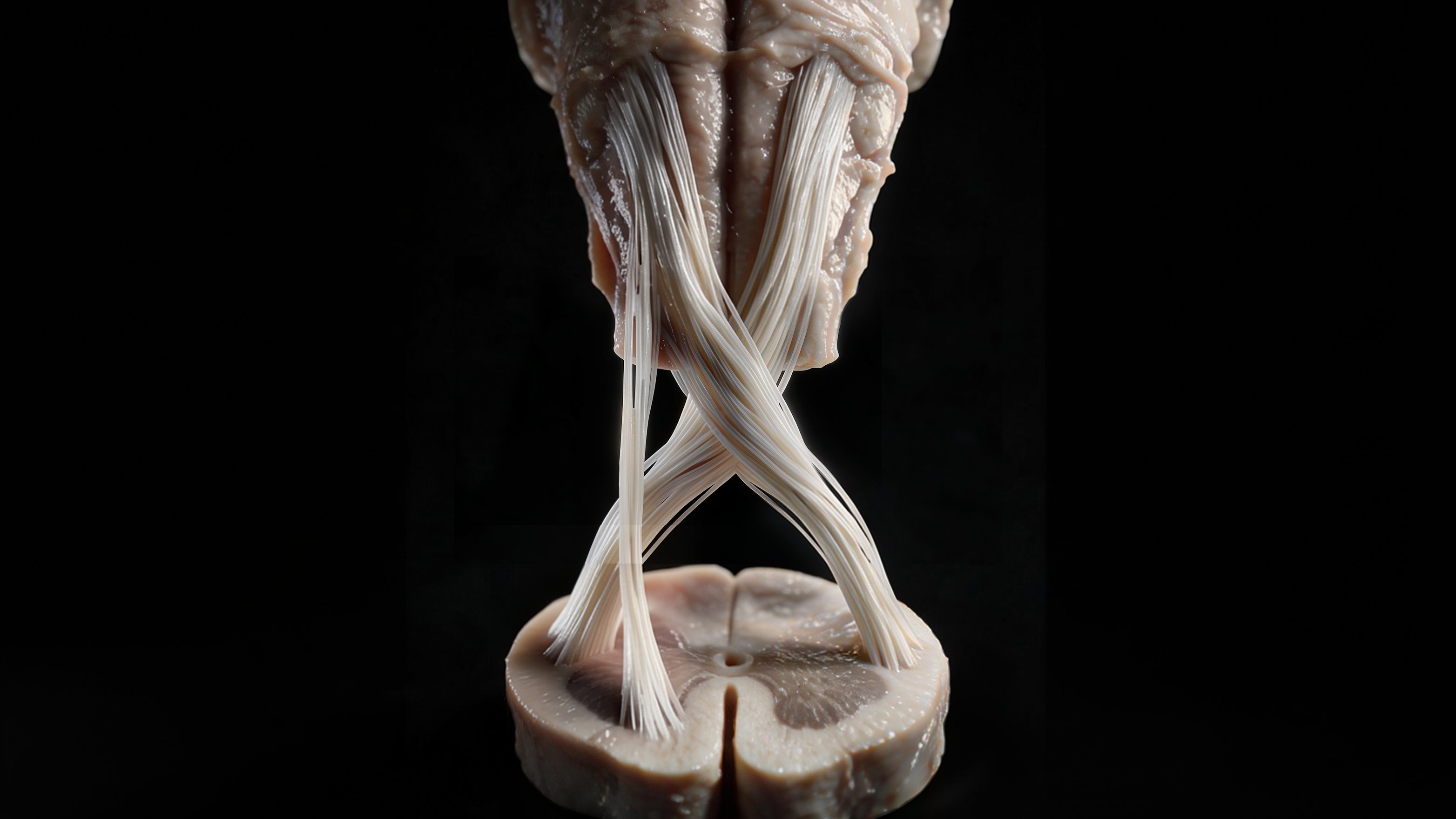
Motor commands leave the cortex, travel down through the brainstem, and at the medulla, roughly 90 percent of the descending fibers physically cross the midline to become the lateral corticospinal tract, continuing down the opposite side of the spinal cord from where they started. The smaller remainder never crosses here, continuing straight down as the anterior corticospinal tract. Both terminate in white matter only, never in gray matter, never near the dorsal horn.
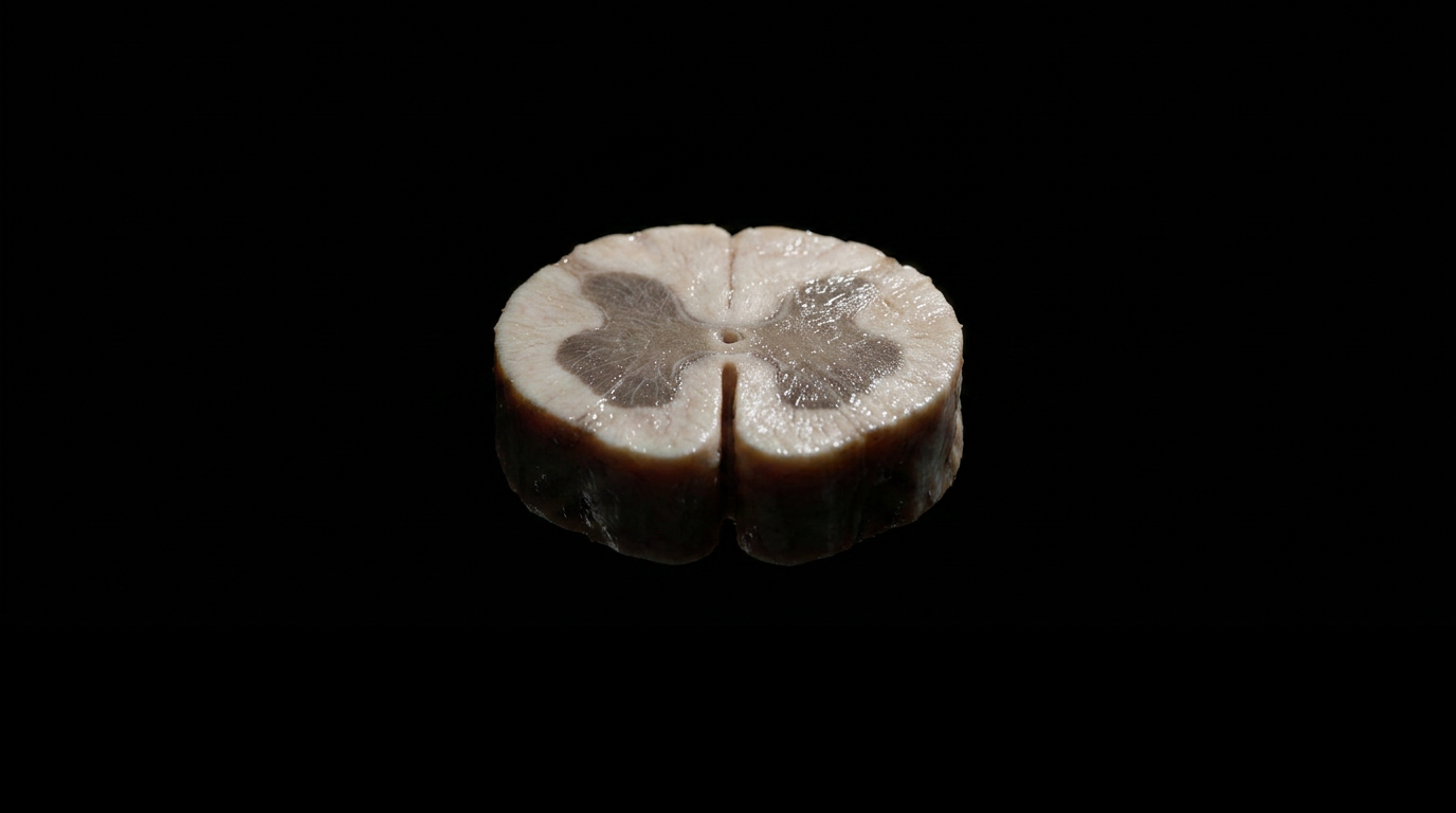
Sensory tracts cross too, just at different levels: the spinothalamic tract crosses low, near the spinal cord itself, so by the time it reaches the medulla it's already carrying signal from the opposite side of the body. The spinal trigeminal tract, carrying facial sensation, hasn't crossed yet at this level. That's why one stroke in one small patch of tissue produces a face that's numb on the same side and a body that's numb on the opposite side. Two tracts, two different crossing points, one lesion.
A previously healthy 40-year-old man finished a high-intensity leg workout, heavy squats and leg presses, holding repeated neck extension and rotation under load. About thirty minutes later: sudden headache on one side, vomiting, trouble swallowing, a hoarse voice, dizziness, and numbness that didn't match any one nerve he could name.
On exam: a partial droop and constricted pupil on one side (an incomplete Horner's triad), severe difficulty swallowing, a hoarse voice, reduced sensation on one side of the face, and reduced pain and temperature sensation on the opposite side of the body. CT angiography found a focal occlusion in the distal segment of the vertebral artery, on the same side as the droop. MRI confirmed an acute infarct in the lateral medulla. Blood work showed a markedly elevated creatine kinase, exertional rhabdomyolysis from the workout itself, a real physical strain on the vessel wall, not a coincidence riding alongside the stroke.
He was started on dual antiplatelet therapy and a high-intensity statin, and began intensive rehab. At discharge: stable, meaningfully improved, with some residual swallowing and voice difficulty still resolving under continued therapy. Source: peer-reviewed case report, "Wallenberg Syndrome After Leg Day Training," PMC, 2026.
Localizing a brainstem lesion isn't pattern memorization. It's roll call. Every finding either belongs to a fixed nuclear address or it doesn't, and the addresses that don't answer are just as diagnostic as the ones that do. A young, healthy man walks out of a gym with a stroke because a vertebral artery, stretched and torn under repeated neck extension and load, stopped feeding one small wedge of his brainstem. The vestibular nuclei misfired, the shared nucleus for cranial nerves IX and X went dark together, the sympathetic fibers dropped out, and the two sensory tracts, crossing at two different levels, produced a numbness pattern that looks scrambled until you know where each one crosses. Nothing here was random. Every symptom had an address, and the address that never answered, the silence from cranial nerves XI and XII, told the story just as clearly as the symptoms that did.
This case went from documented patient to fully rendered illustration in real time. New case studies publish the same way: the mechanism, the imaging, the outcome, live. Get notified the moment the next one goes up.
No spam. Unsubscribe anytime. New case study alerts only.
You're on the list. We'll notify you when the next case goes live.
You're set. We'll send the full write-up to your inbox shortly.