Fat Necrosis

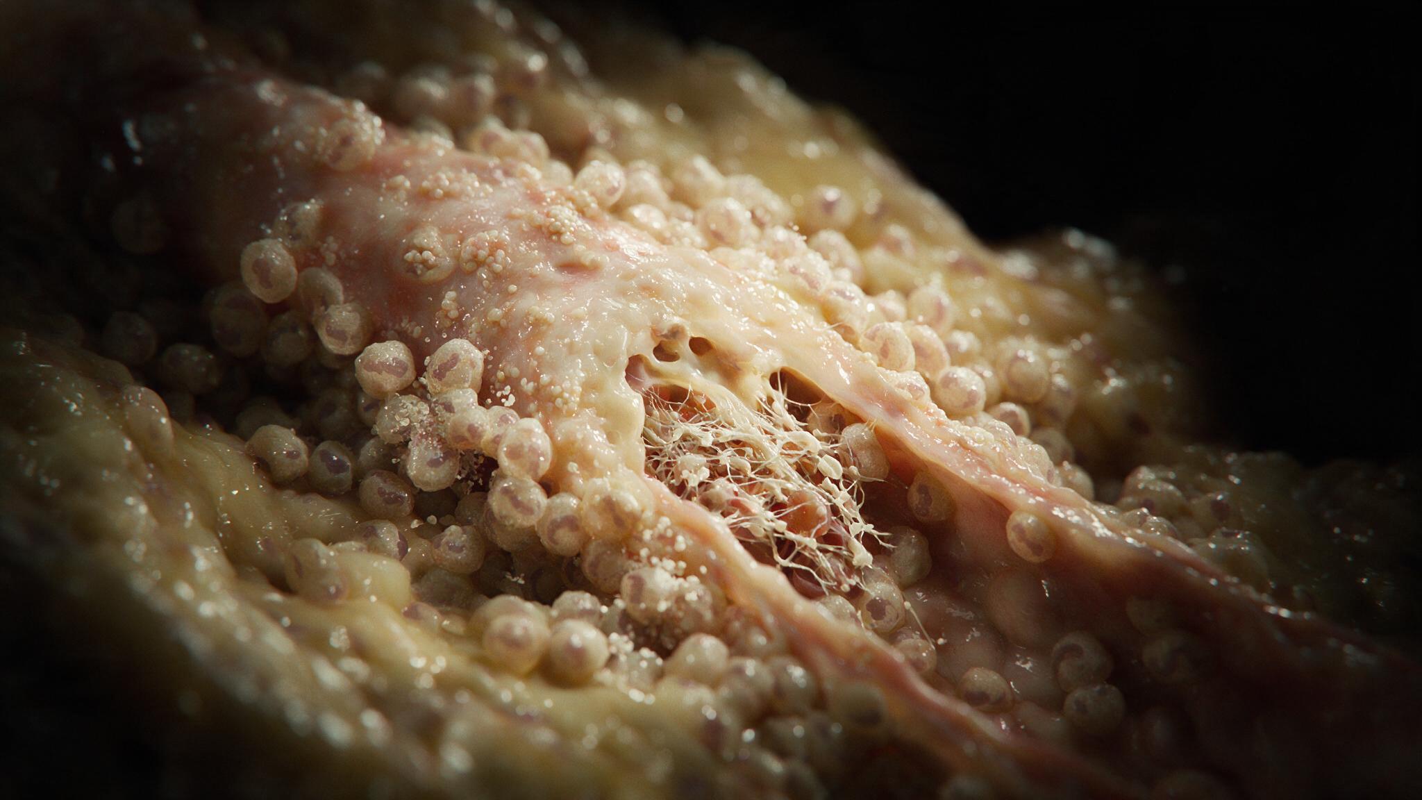
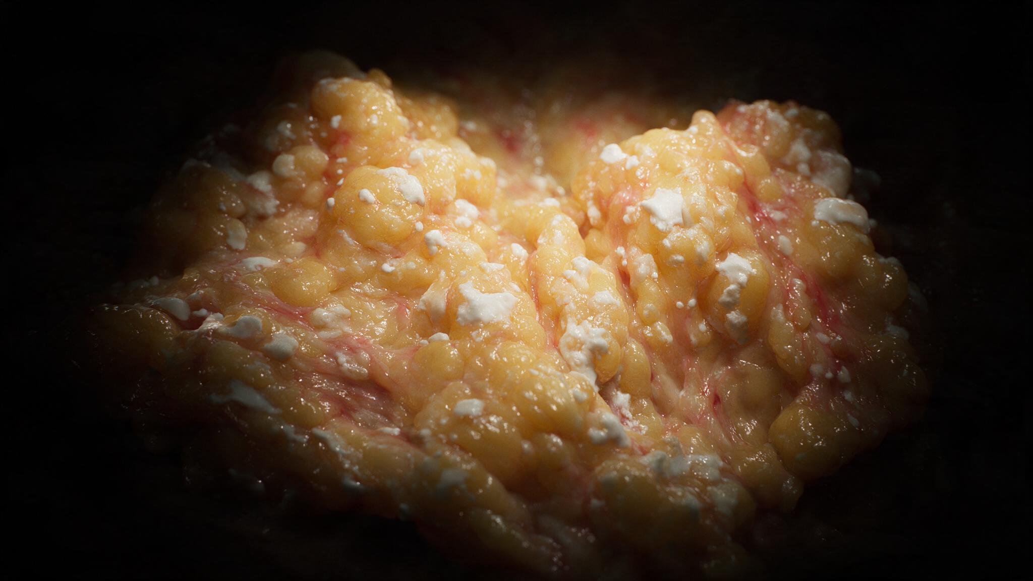
Open the abdomen during acute pancreatitis and the omentum tells you what happened before you even see the pancreas. Small chalky-white plaques, the color of candle wax, scattered across the fat. No other necrosis looks like this.

There are two ways to get there. Pancreatic lipase digesting fat from the inside, or a blunt injury tearing it open by force. Same white lesion, two completely different stories, and the second one is the case most clinicians actually see.

Case TypeClinical Illustration
FocusPathology
Year2026
Operative view of the greater omentum with scattered chalky-white saponified fat necrosis plaques. Pancreatic lipase has digested triglycerides, and free fatty acids have bound calcium to form insoluble soaps
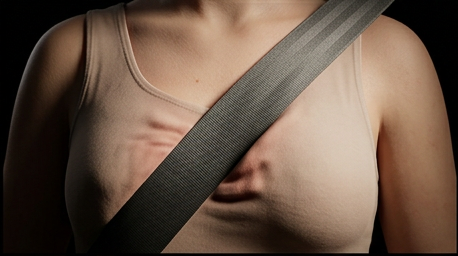
The moment of impact: a diagonal seatbelt strap pressed hard across a woman's chest, skin and soft tissue visibly compressed under sudden mechanical force

Progression

Enzymatic: Lipase Digests Fat From the Inside

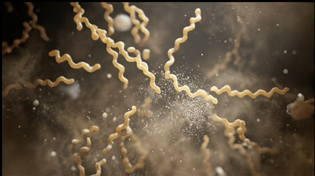
Pancreatic lipase leaks into the peritoneal cavity during acute pancreatitis and splits triglycerides into free fatty acids and glycerol. Those fatty acids grab calcium out of the interstitial fluid and precipitate as an insoluble soap, right inside the dying adipocyte.
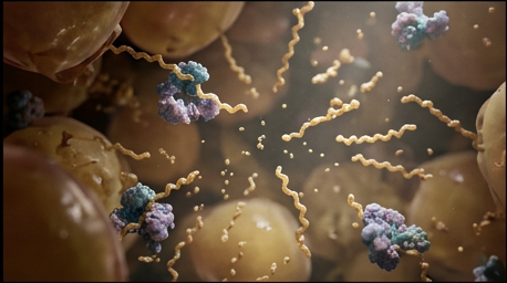
Extreme macro of pancreatic lipase enzyme molecules actively cleaving triglyceride bonds within the fat lobule, releasing free fatty acid chains and glycerol into the surrounding fluid
Figure 1
Lipase cleaving triglyceride bonds inside the fat lobule
Free fatty acid chains physically bonding with calcium ions from interstitial fluid, fine crystalline calcium-soap deposits forming within the fatty debris
Figure 2
Free fatty acids binding calcium to form insoluble soap

This is why severe pancreatitis can cause hypocalcemia: calcium gets consumed by saponification faster than the body replaces it, and hypoalbuminemia, PTH resistance, and impaired vitamin D metabolism pile on from there.

Other white lesions on the omentum
  • TB peritonitis: miliary white tubercles, not soft plaques
  • Metastatic carcinoma: firm nodules, "omental cake"
  • Pseudomyxoma peritonei: gelatinous mucinous deposits
  • Calcified nodes: old granulomatous infection
  • Talc: foreign-body reaction from surgical gloves

Traumatic: Direct Rupture, No Enzyme Required

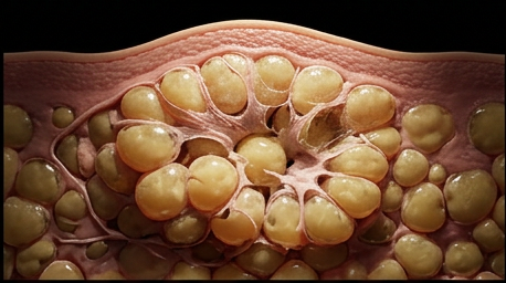
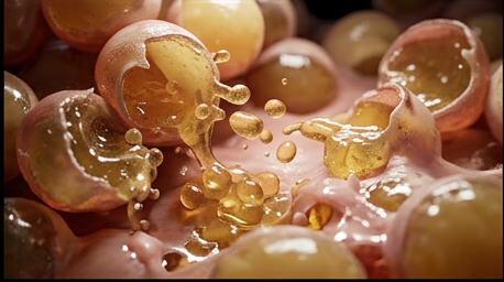
A blunt impact, a seatbelt slamming across the breast, a hard fall onto the buttock, physically shears the adipocyte membrane open. No lipase. No pancreas. The cell dies on the spot from mechanical force, not digestion.
The moment of impact: a diagonal seatbelt strap pressed hard across a woman's chest, skin and soft tissue visibly compressed under sudden mechanical force
Figure 3
Seatbelt compression at the moment of impact
Macro cutaway just beneath the skin at the point of compression, a cluster of adipocytes physically crushed and rupturing under shear force, thin cell membranes tearing
Figure 4
Adipocytes crushed and rupturing under shear force
Extreme macro at the moment of membrane rupture, glistening triglyceride lipid droplets spilling out of burst adipocytes into the surrounding interstitial space
Figure 5
Triglyceride droplets spilling from burst adipocytes

From there the body runs the same cleanup either way: foam cells clear the debris, giant cells wall off the bigger deposits, and the site slowly organizes into fibrosis, sometimes calcifying at the rim.
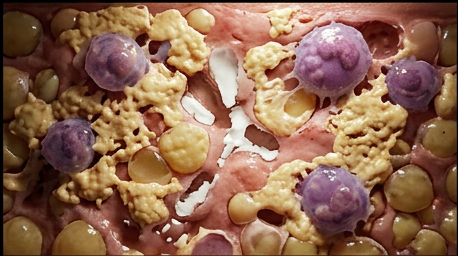
Foamy lipid-laden macrophages and multinucleated giant cells surrounding pools of cholesterol debris, real cellular cleanup activity
Figure 6
Foam cells and giant cells clearing lipid debris
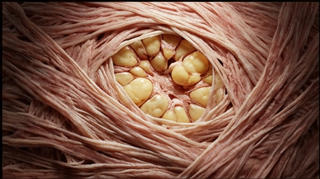
Dense fibrous connective tissue physically encapsulating the lesion from the outside in, collagen fibers wrapping and walling off the calcified fatty core
Figure 7
Collagen fibers walling off the calcified core

Documented Case: The Lump That Waited Twelve Years

A 51-year-old woman found a painless lump in her left breast weeks after a car accident. The seatbelt had bruised that same breast. The lump sat stable for over a decade, until it started growing and she came in for a biopsy. The result: fat necrosis. No cancer. Just the fibrosed, calcified remnant of a seatbelt injury from ten years earlier. Source: PMC case report, ruptured giant expanding breast oil cyst.
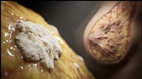
Depth-split frame: a chalky-white omental plaque in extreme macro foreground, a fibrosed calcified breast lesion in cross-section soft-focus background
Figure 8
Omental plaque and breast lesion, same process, two sites

This is the trap. A real injury, a gap long enough that the patient nearly forgot the cause, and an imaging picture that looks exactly like cancer until someone samples the tissue. That gap is why "fat necrosis, no evidence of malignancy" is one of the best phrases you can read in a pathology report.

Outcome

Two roads, same lesion. In the omentum, lipase digests fat into a chalky-white soap. In the breast, a blunt injury tears the fat open and the body runs the same fibrotic cleanup anyway, no enzyme needed. Most calcified deposits stay stable, but not all: some enlarge, some infect, which is why a growing lump years after an old injury still gets imaged and, when uncertain, biopsied rather than waved off by history alone. Both mark the site of an old insult the way a flood leaves a high-water line: the omental plaque for a pancreas that leaked, the breast lump for a seatbelt that closed too hard, too fast.
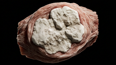
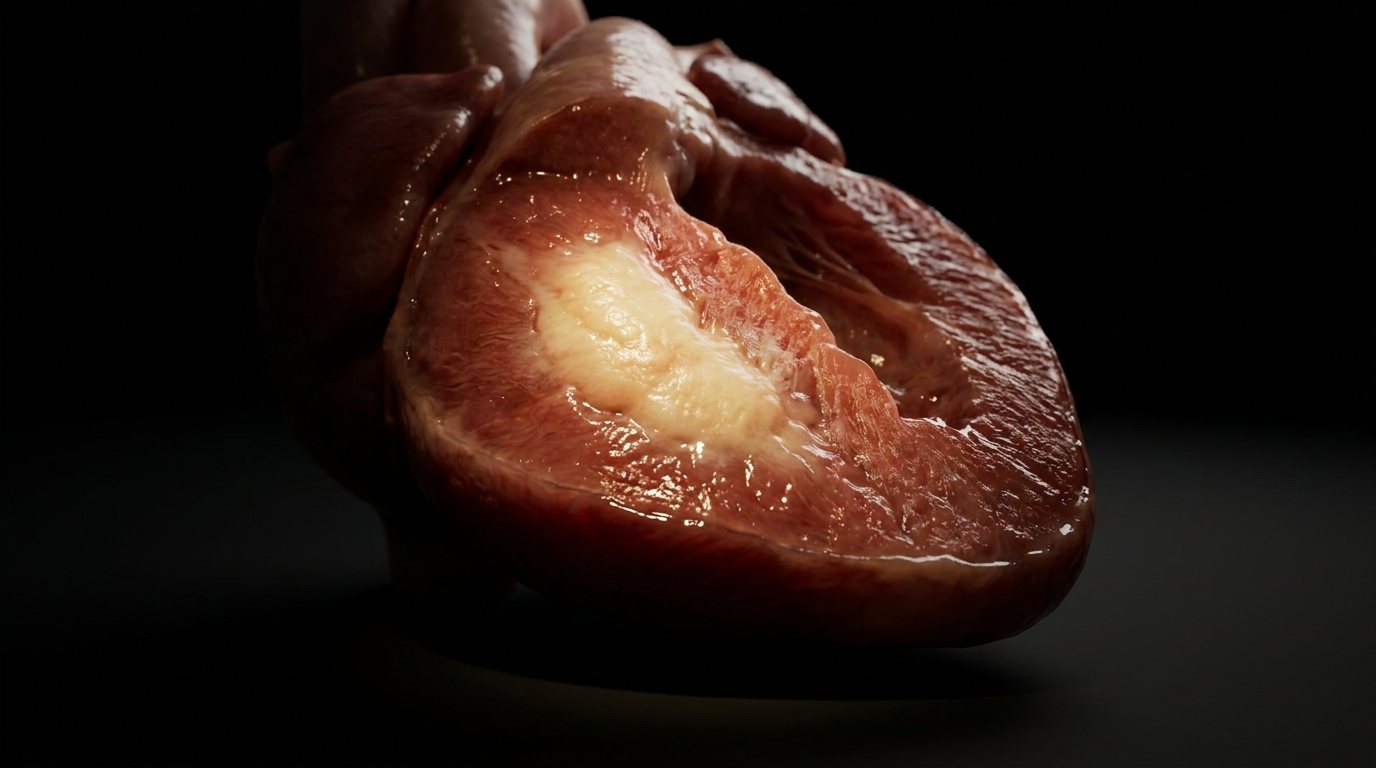
The final stable lesion years later: a firm, chalky-white, irregularly calcified nodule in cutaway cross-section, encapsulated in fibrous tissue
Figure 9
The final stable lesion, years after the original injury

Other work

Arthus Reaction

Arthus Reaction

Medical Illustration

Coagulative Necrosis

Coagulative Necrosis

Medical Illustration

Want to learn about the human body
from the inside?

steven.oppong@gmail.com