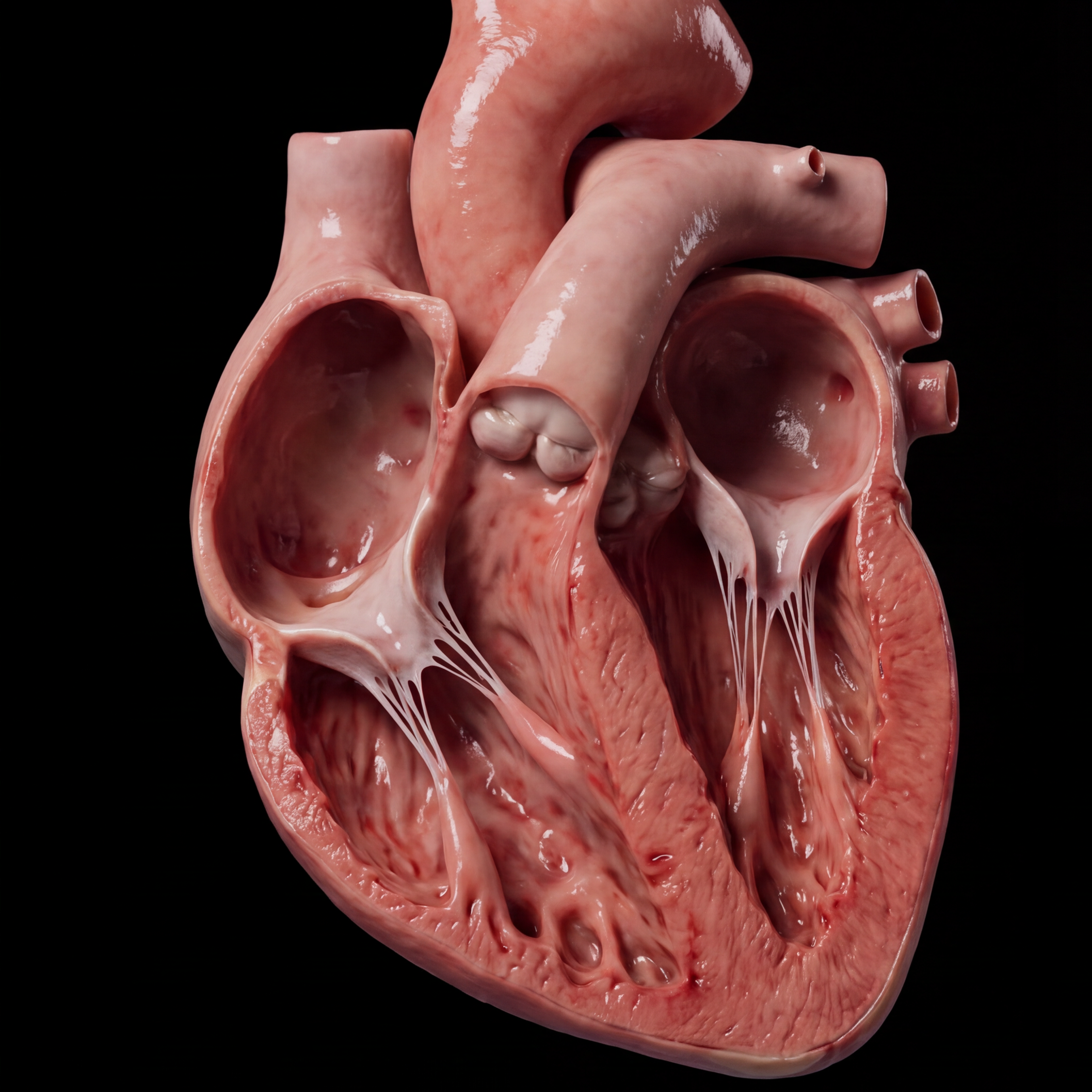
A pulmonary embolism begins quietly, often in the deep veins of the leg, where blood stagnates and a clot takes shape. By the time it reaches the lungs, it has traveled through the right side of the heart and lodged in a pulmonary artery, setting off a cascade of hemodynamic strain that can turn fatal within hours.
This case study traces the pathophysiology of venous thromboembolism from Virchow's triad through right ventricular failure. Each panel was built to make the invisible sequence visible: stasis, hypercoagulability, endothelial injury, clot propagation, and the final mechanical standoff between a struggling right ventricle and an obstructed pulmonary circuit.
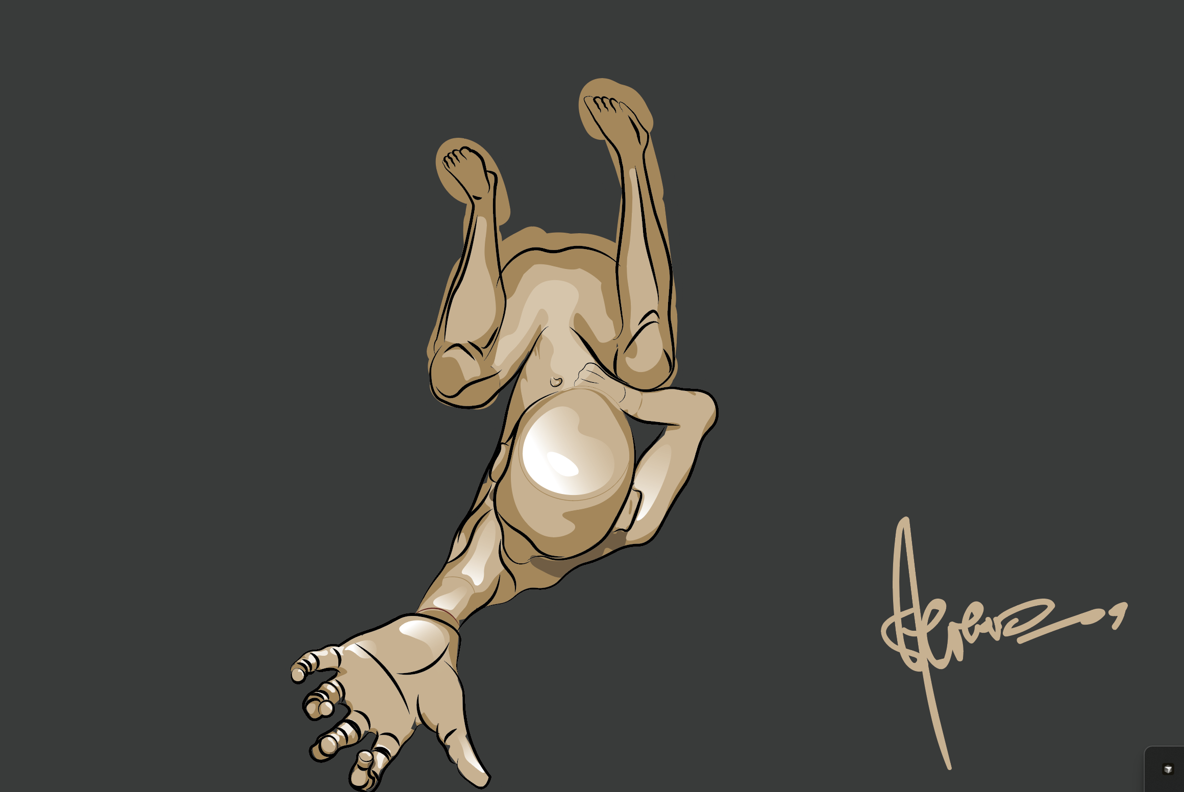
The story of every thrombus begins with three converging forces. Stasis slows blood flow in the deep veins, allowing clotting factors to accumulate. Hypercoagulability tilts the balance toward fibrin formation. Endothelial injury exposes the subendothelial matrix, triggering platelet adhesion and the coagulation cascade. Together, these three conditions create the environment a clot needs to nucleate and grow, often silently, in the deep veins of the lower limb.
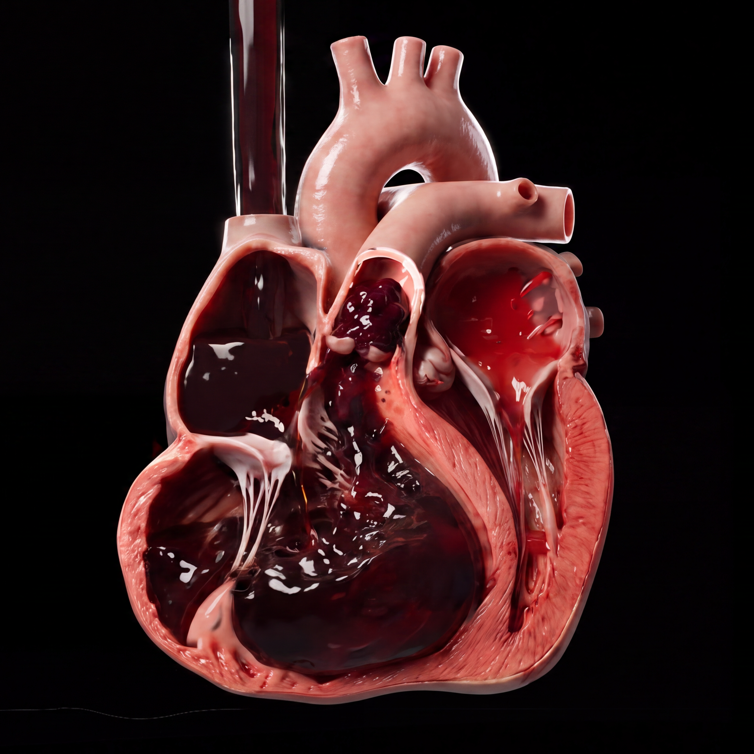
Once a fragment of thrombus breaks free, it travels as an embolus through the venous system, into the right atrium, through the right ventricle, and out into the pulmonary circulation. There it may lodge anywhere in the pulmonary circuit. The obstruction is mechanical: blood cannot reach the alveolar capillary beds for gas exchange. But the deeper problem is hemodynamic: the right ventricle, a thin-walled chamber built for low-pressure flow, now pumps against a suddenly elevated pulmonary vascular resistance. It strains, dilates, and begins to fail.
Right-heart strain is not just a complication; it is the message the body sends when the pulmonary circuit is under siege. On ECG, the strain pattern appears. On echocardiography, the right ventricle bulges into the left, reducing cardiac output. Treatment with thrombolytics like alteplase dissolves the fibrin meshwork, restoring flow. The strain recedes. The right ventricle, if it held on long enough, recovers. What looked like mechanical obstruction was, at every step, a signal waiting to be read.
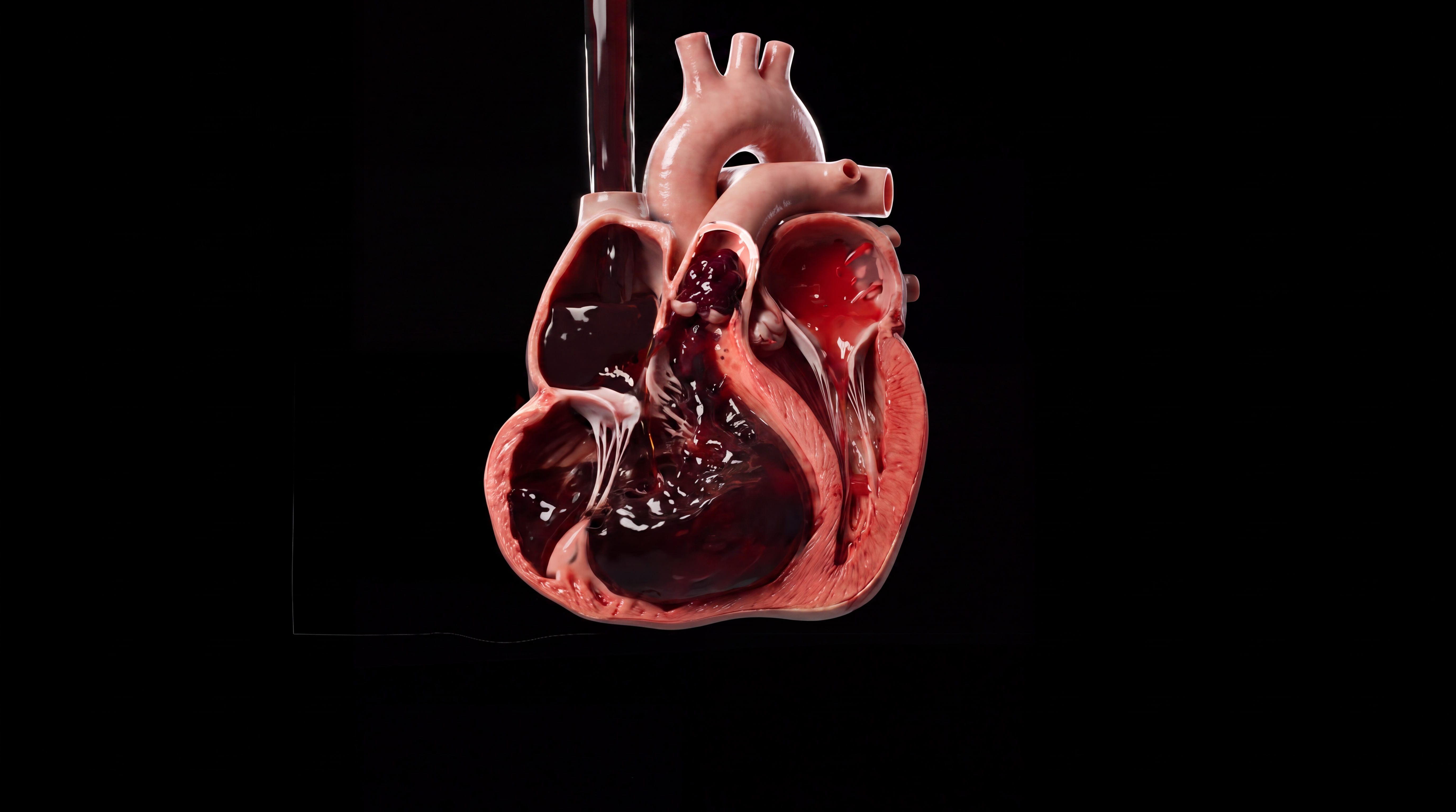
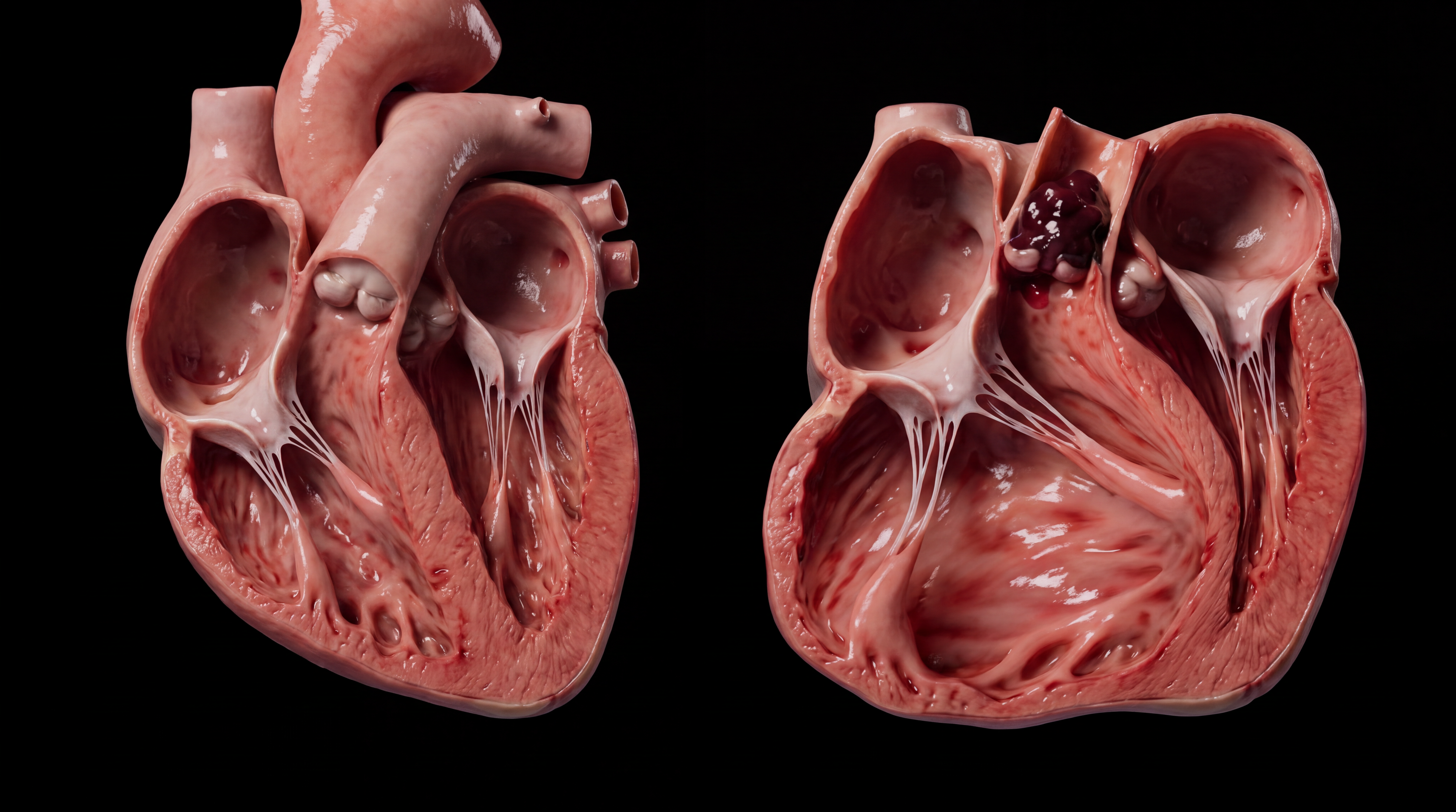
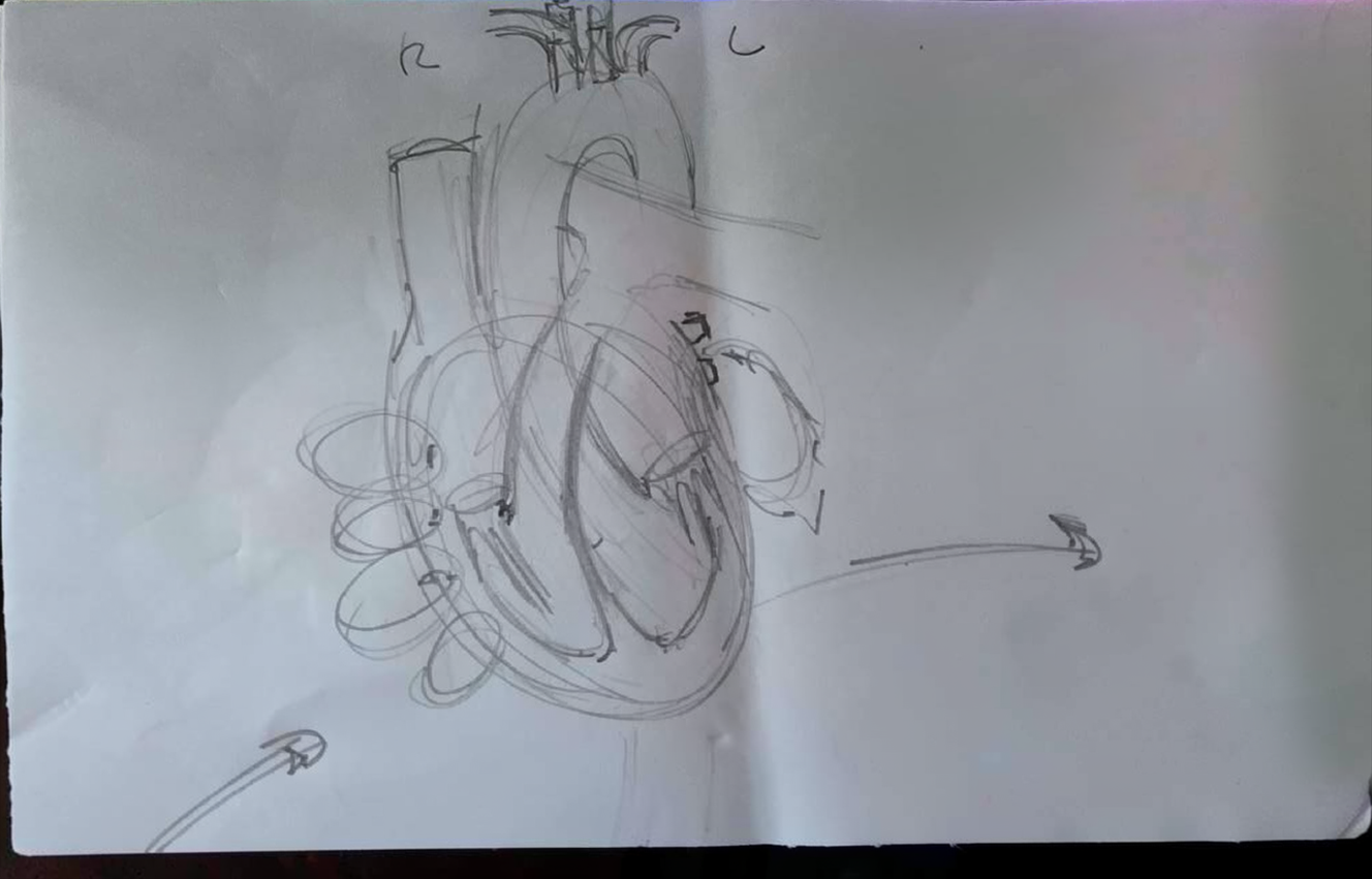
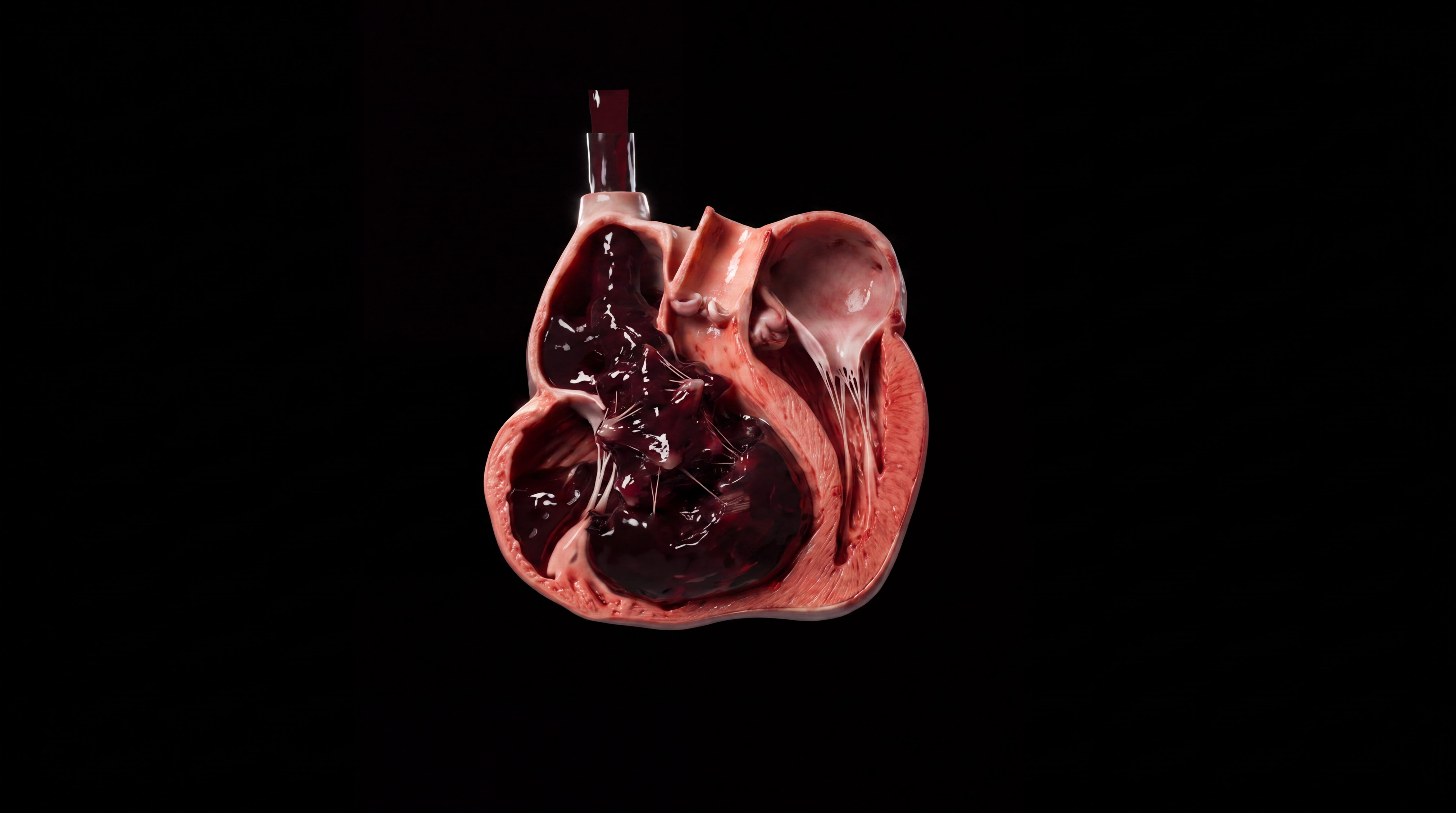
The patient received alteplase within the window. The clot dissolved. Pulmonary artery pressure fell, and oxygen saturation climbed back toward normal. The right ventricle, relieved of its burden, returned to its natural geometry over the following days. This is the earned ending: a system pushed to the edge of failure, pulled back by timely intervention. The illustrations in this series were built to capture that arc — from silent clot formation to the final relief of restored flow — because understanding the mechanism is what makes the ending legible.
You're set. We'll send the full write-up to your inbox shortly.