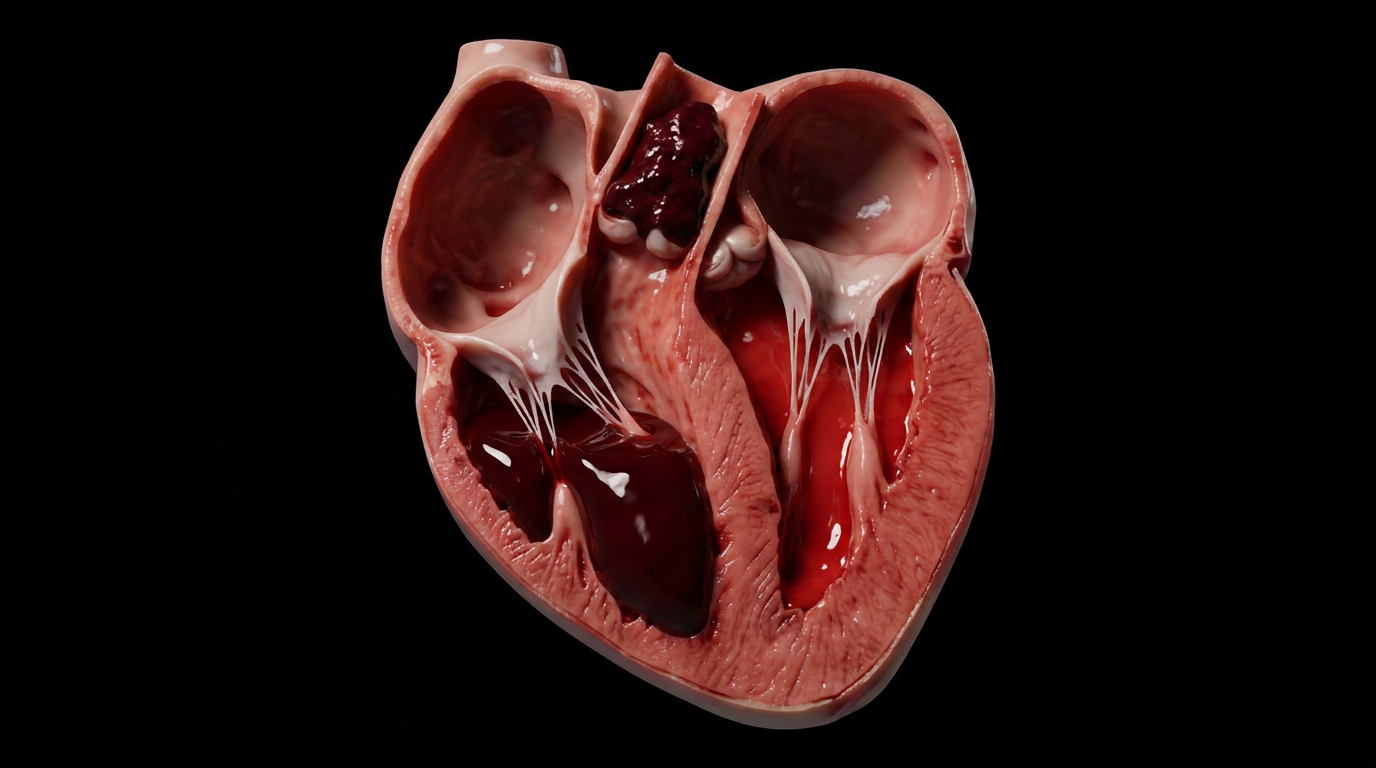
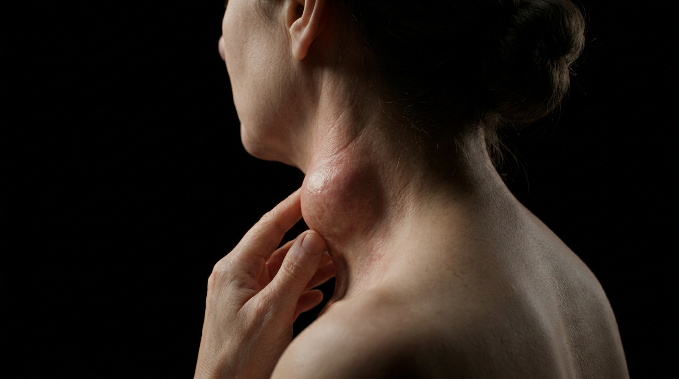
The neck swells. No heat, no redness, no tenderness. Just a slow, quiet enlargement of the cervical lymph nodes that the patient dismisses for weeks. When it finally breaks through the skin, what drains is not pus in the usual sense: it is caseous necrosis made liquid, the debris of a granulomatous war fought and lost inside a lymph node.
Scrofula is cervical tuberculous lymphadenitis, the most common form of extrapulmonary TB. The bacilli seed the cervical nodes, multiply inside macrophages, and provoke the body's most elaborate containment strategy: the granuloma. When that strategy fails, what was walled off becomes a cold abscess, and the disease announces itself not with fever but with a fistula.
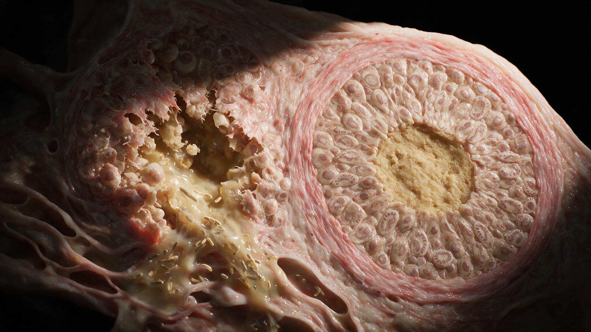
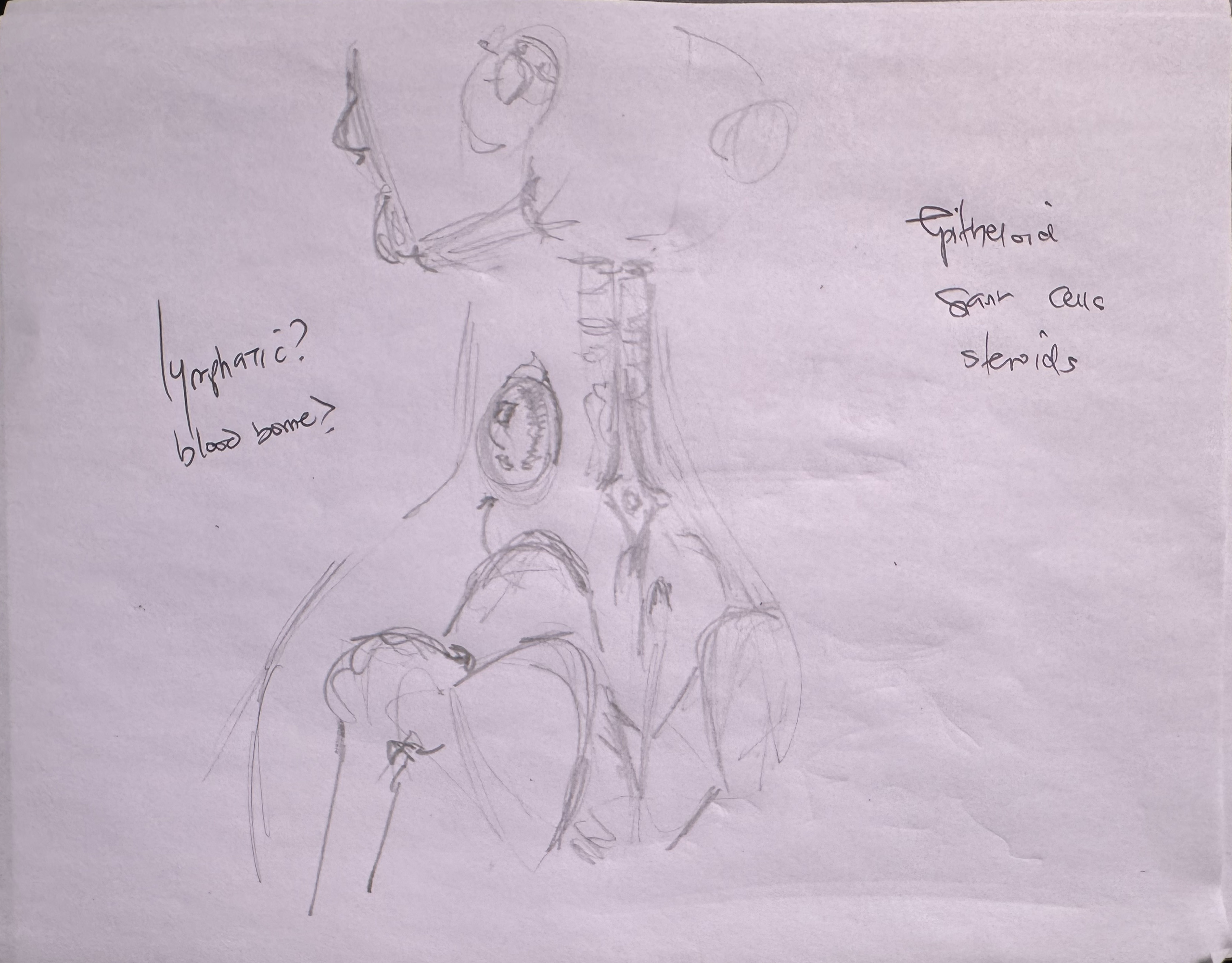
Mycobacterium tuberculosis reaches the cervical lymph nodes not by draining the lungs but through the bloodstream. Hematogenous dissemination from a pulmonary focus seeds bacilli into the node. Macrophages engulf them but cannot kill them. In response, the immune system builds a granuloma: a spherical fortress of epithelioid histiocytes, Langhans giant cells, and a peripheral rim of lymphocytes and fibroblasts. The center undergoes caseous necrosis: a dry, cheese-like consolidation of dead cells held in place by the collagen rim. This is containment. The bacilli are walled off, and the patient is asymptomatic. The disease is present but silent.
Containment is not permanent. The caseous core begins to liquefy. The epithelioid sheet that lined the granuloma wall detaches. The collagen rim weakens, cracks, and the liquefied necrotic debris, now teeming with bacilli, spills out of the granuloma into the surrounding nodal tissue. This is the tipping point. What was a contained infection becomes an expanding one. The node enlarges. The overlying skin stretches. The mass is still painless because the process is indolent, not acute. There is no neutrophil-driven inflammation, no rubor, no calor. Hence the name: cold abscess.
The liquefied caseous material tracks through the node, breaches the capsule, and follows tissue planes toward the skin. Bacilli also breach nearby vessel walls, opening a route for further hematogenous spread. The skin over the node thins, reddens slightly, and finally ruptures, producing a sinus tract that drains thin, cheesy material. This is scrofula in its classic presentation: a chronic draining fistula in the neck, discharging the debris of a years-long immune containment battle. The bacilli that spill out can seed new nodes, skin, or return to the lungs. The cold abscess is not the end; it is the beginning of the next cycle.
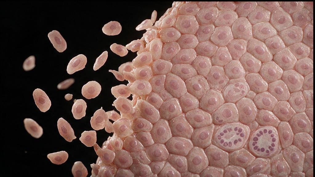
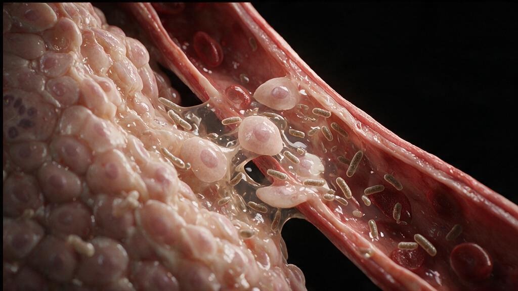
Treatment is medical, not surgical. The standard four-drug TB regimen (rifampin, isoniazid, pyrazinamide, ethambutol) penetrates the granuloma and kills the bacilli. The cold abscess heals, the fistula closes, and the node slowly fibroses and calcifies over months. Incision and drainage is avoided where possible: it creates a chronically draining sinus that is harder to heal than the disease itself. The illustrations in this series trace the arc from hematogenous seeding to fistula formation, because understanding why the abscess is cold, why the node is painless, and why the granuloma must fail before the disease declares itself is what makes the clinical presentation legible.
You're set. We'll send the full write-up to your inbox shortly.