OB-GYN Textbook Plates

A set of obstetric and gynecologic anatomical illustrations commissioned for a medical textbook. The plates follow the clinical arc from fetal anatomy through labor assessment, lie and presentation, pelvic brim anatomy, and perineal injury classification.

Each plate was drawn to serve two audiences simultaneously: the student learning anatomy for the first time, and the clinician who needs to reference specific landmarks under pressure. The illustrations prioritize clarity of spatial relationships over decorative detail, because in obstetrics, millimeters at the pelvic brim determine management decisions.

Publication Partograph
Focus Obstetrics & Gynecology
Plates 10 illustrations

Fetal Anatomy & Skull Moulding

The fetal skull is not a rigid structure. The bones of the calvarium are separated by sutures and fontanelles, allowing the head to mould and adapt as it passes through the birth canal. These plates show the process of moulding and the clinical consequences when compression exceeds the scalp's tolerance.
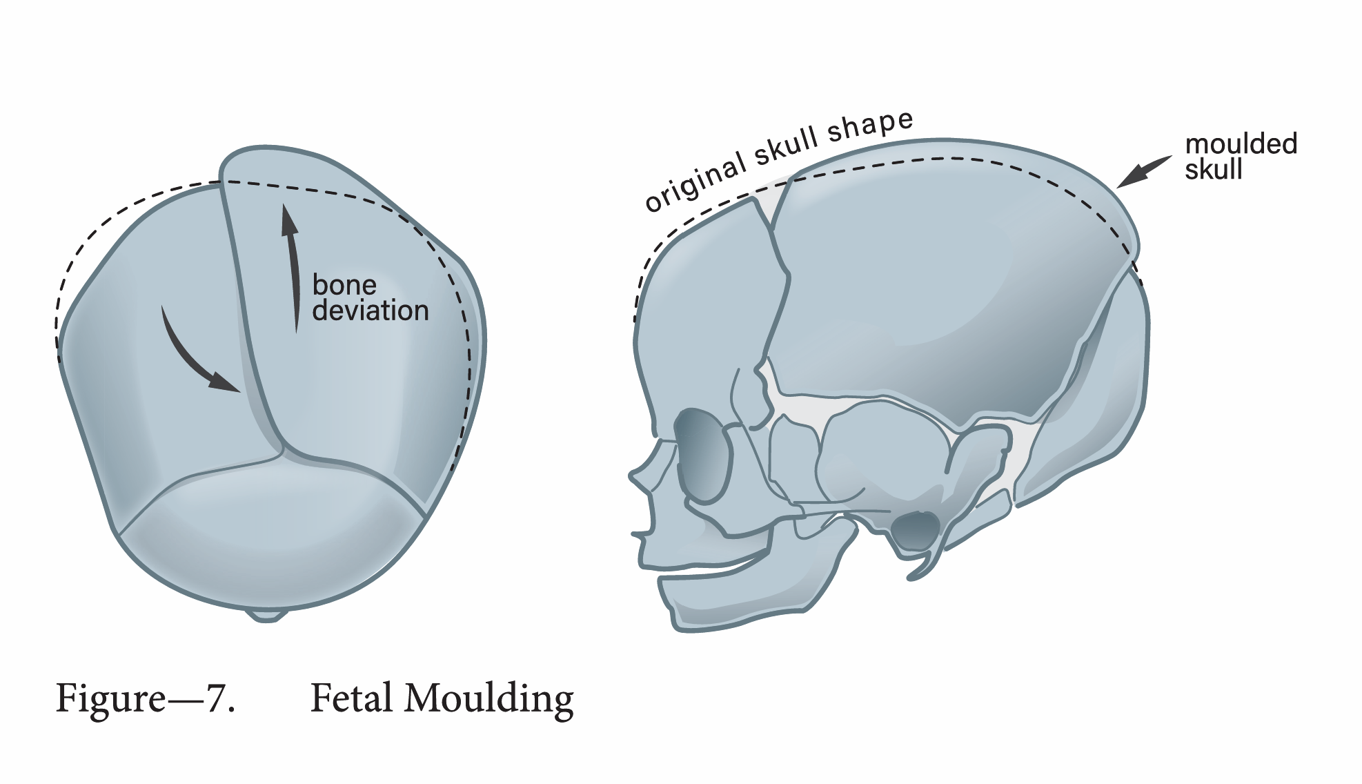
Fetal moulding of the skull
Figure 7
Fetal Moulding — adaptive compression of the fetal skull during descent through the maternal pelvis
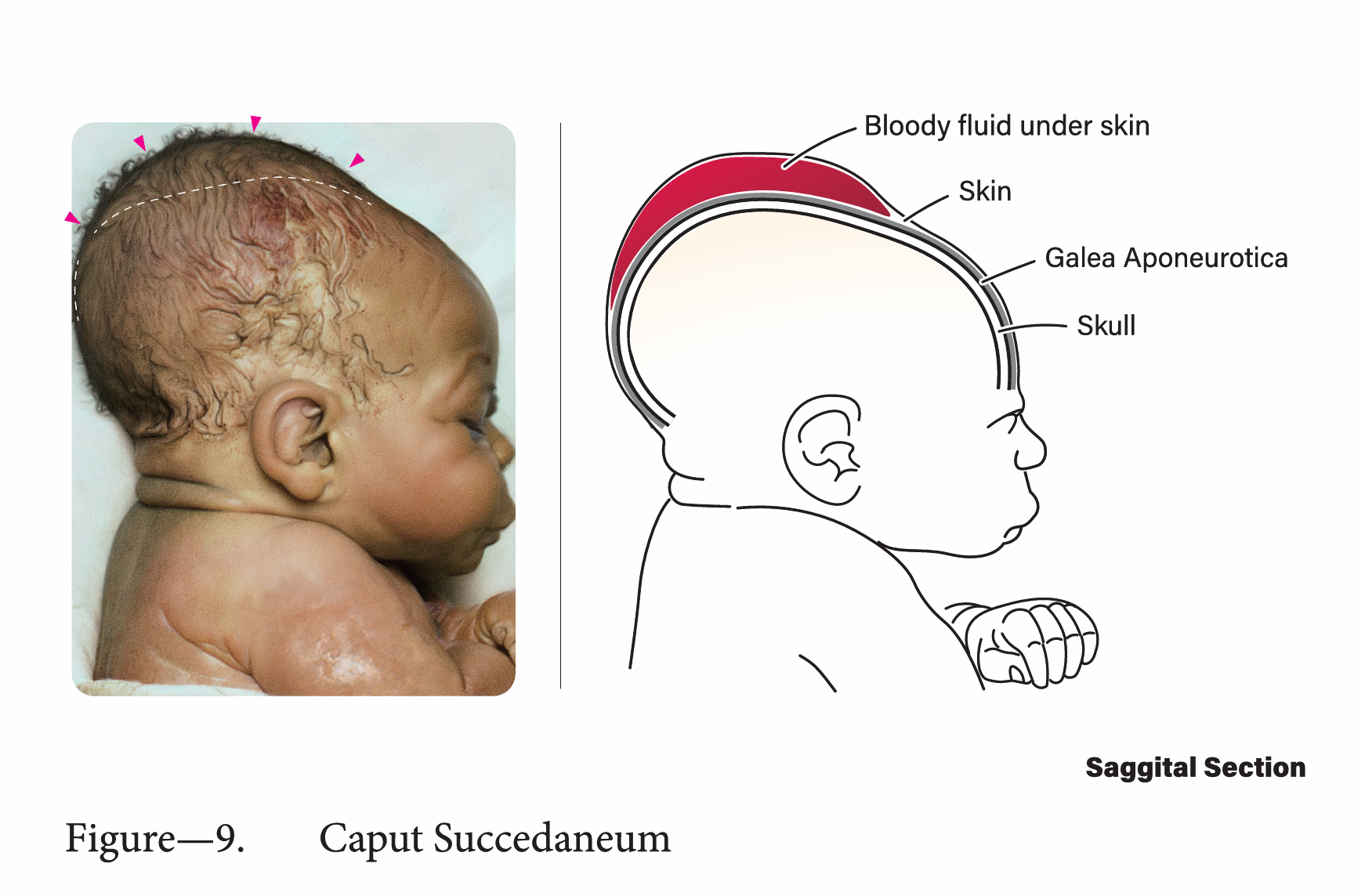
Caput succedaneum and neonatal scalp injury
Figure 9
Caput Succedaneum — serosanguinous swelling of the neonatal scalp resulting from sustained pressure against the cervix
Labor Assessment: Descent & Pelvic Brim

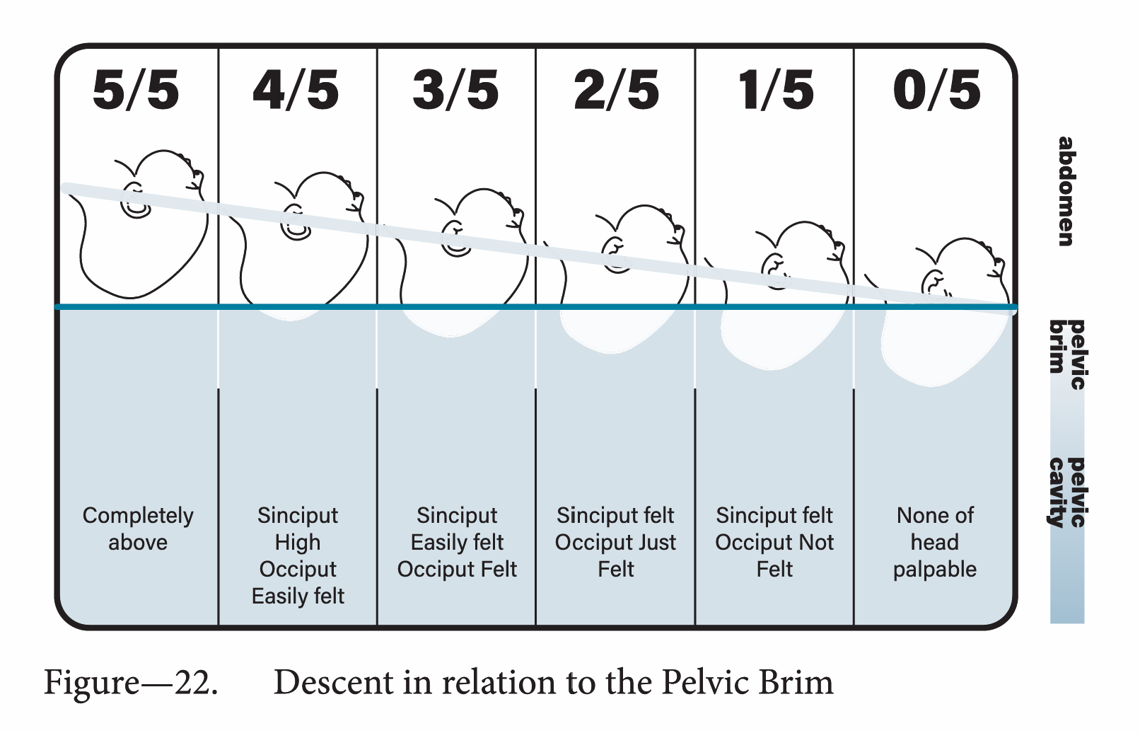
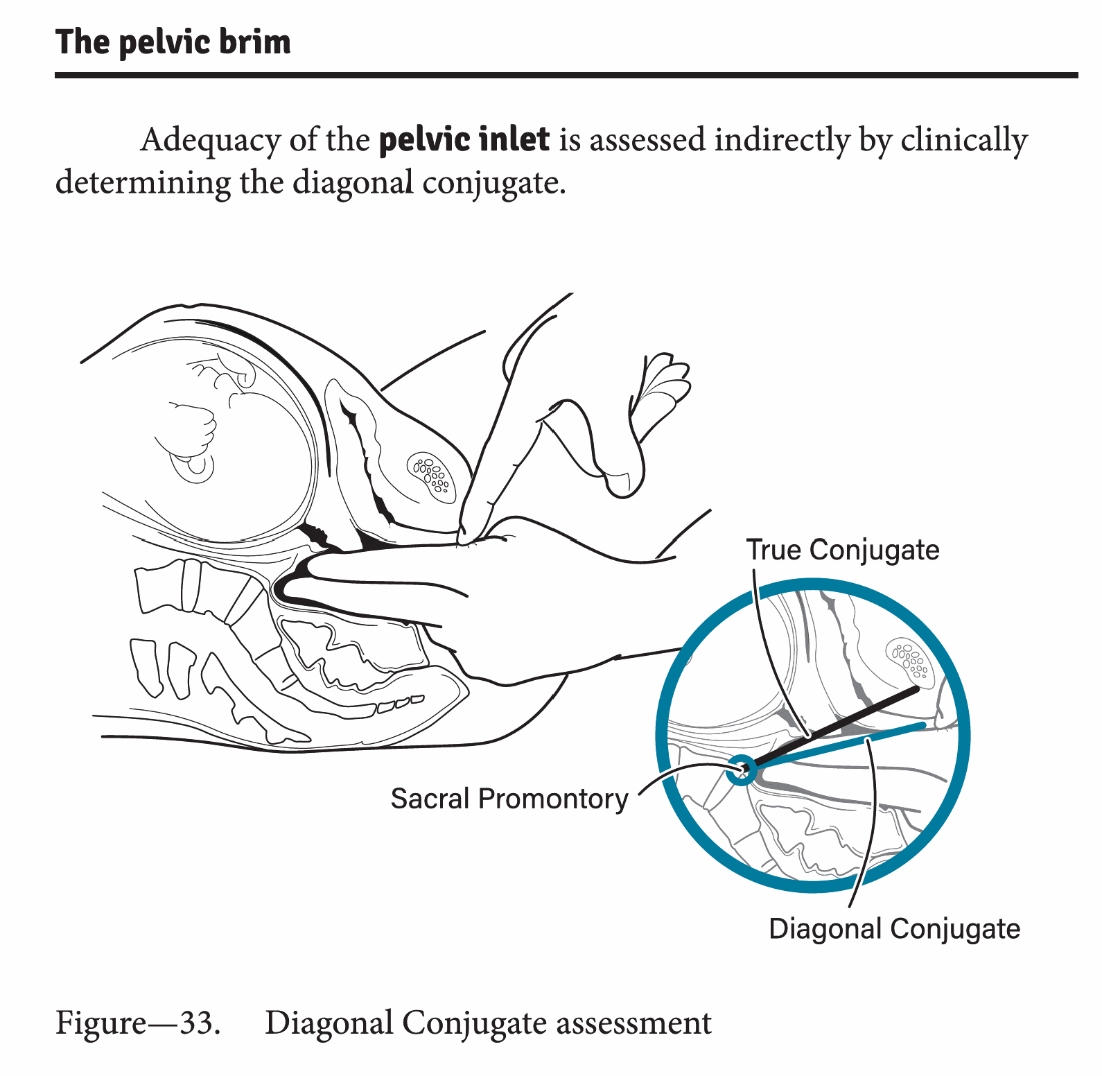
The first determination in every labor assessment is whether the fetal head can navigate the pelvic inlet. These plates map descent in relation to the pelvic brim, and illustrate the conjugate diameters used to assess pelvic adequacy, landmarks that cannot be visualized directly in a living patient but must be understood three-dimensionally to interpret clinical findings.

Descents in relation to the pelvic brim
Figure 23
Descent in Relation to the Pelvic Brim — tracking the progressive engagement of the presenting part through the inlet, mid-cavity, and outlet
Pelvic brim conjugate diameters
Figure 39
Diagonal Conjugate Assessment — the true conjugate, diagonal conjugate, and sacral promontory as landmarks for evaluating pelvic adequacy

Fetal Lie & Presentation

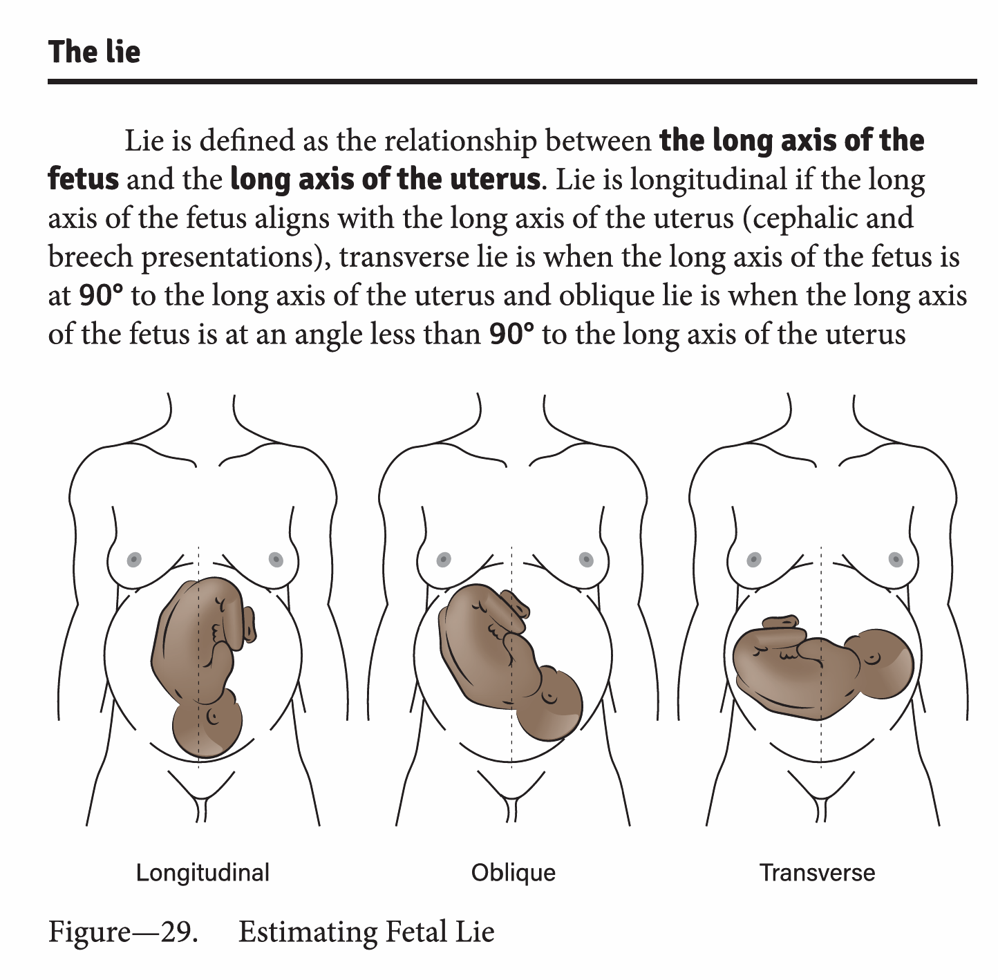
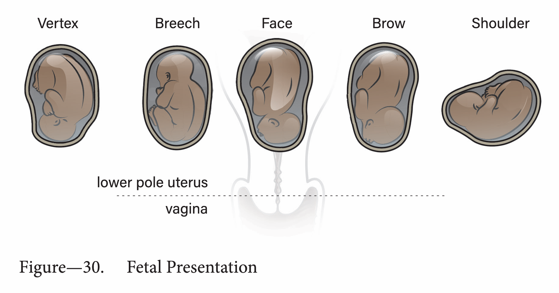
The relationship between the long axis of the fetus and the longitudinal axis of the uterus defines the lie: longitudinal, transverse, or oblique. The presenting part determines management. These plates map both dimensions, providing the visual reference a clinician returns to when palpating a term abdomen.

Fetal lie: longitudinal, transverse, oblique
Figure 29
Fetal Lie — longitudinal, transverse, and oblique positions defined by the relationship of the fetal axis to the maternal uterine axis
Fetal presentation: breech, face, shoulder, vertex
Figure 30
Fetal Presentation — breech, face, shoulder, and vertex presentations, showing the landmark used to identify each variant

Pelvic Injury Classification

Pelvic injuries are graded from first through ninth degree, a classification that compresses a clinical decision tree into a single visual reference. The plate combines anatomical cross-section with the injury grading scale, so the reader can map severity directly onto pelvic landmarks.

Pelvic injury classification by degree
Table
Pelvic Injuries — classification by degree (1st through 9th), mapped onto a cross-sectional view of the female pelvis

Perineal Injury: Intact vs. Degrees of Injury

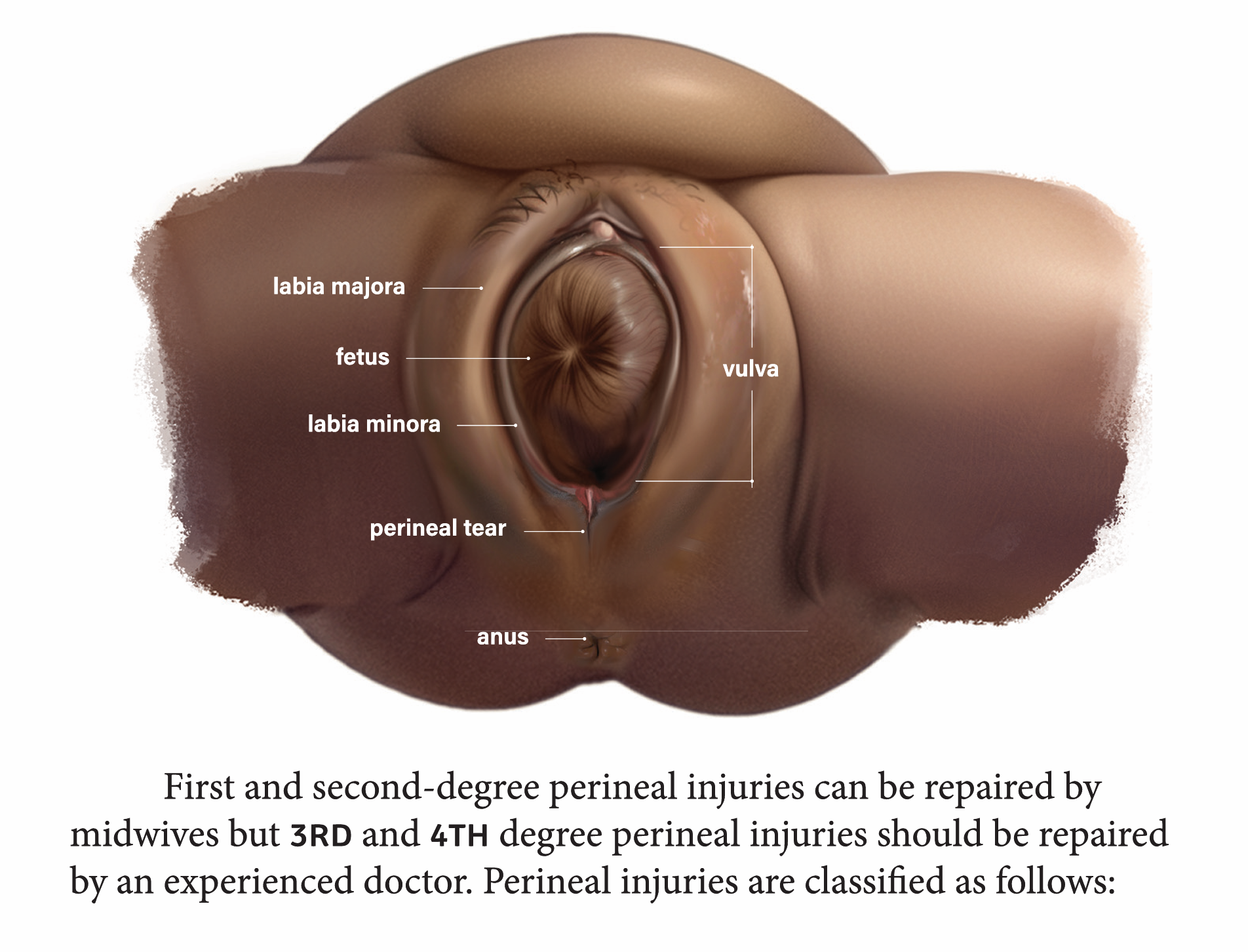
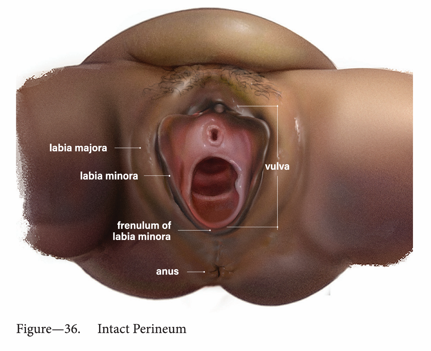
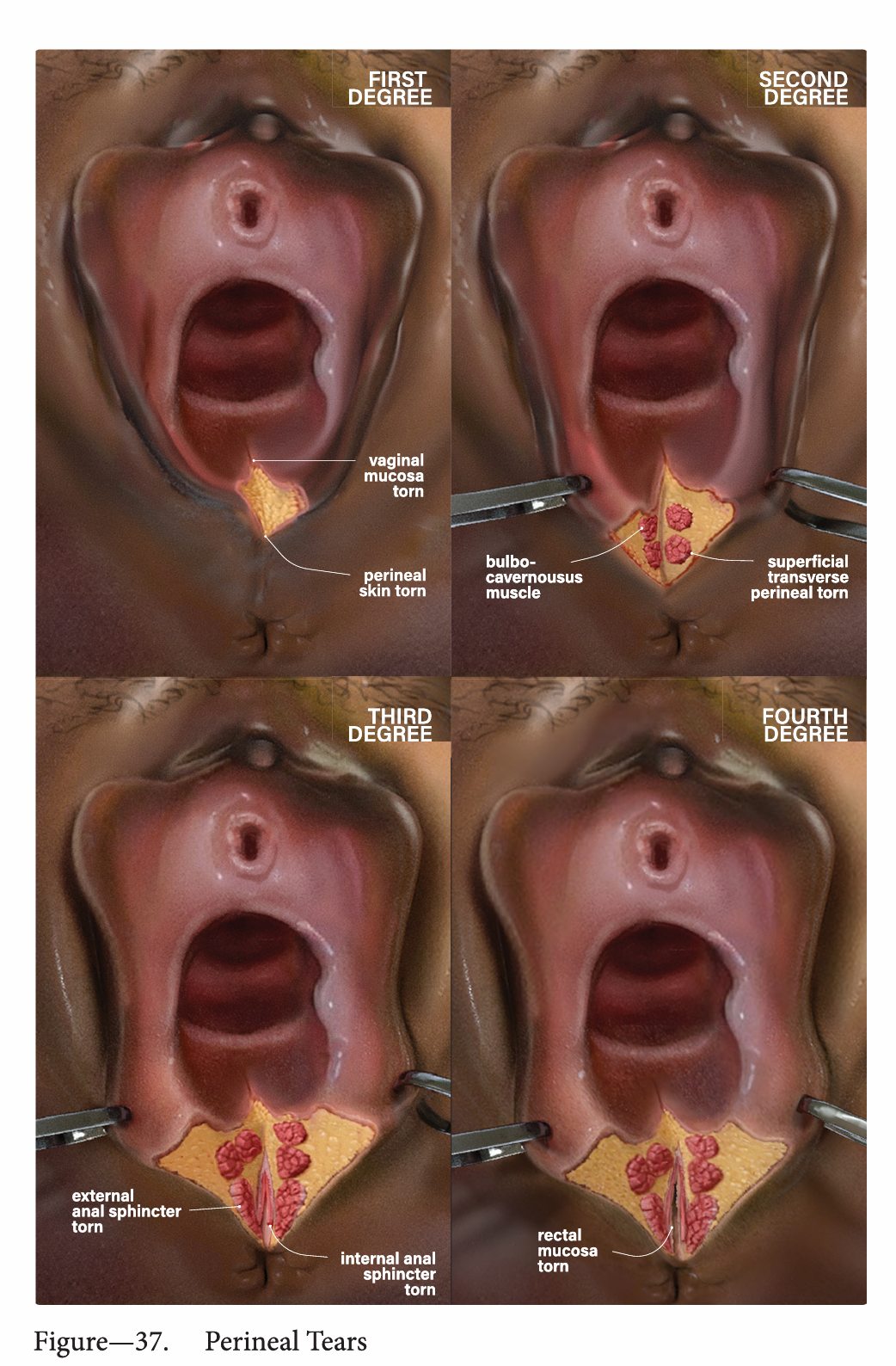
These two plates form a deliberate teaching pair. The intact perineum on the left establishes the baseline anatomy: labia majora, labia minora, vaginal introitus, perineal body, and terminus ani. The injury plate on the right shows the same anatomical view with first- and second-degree perineal injuries, using the same landmarks to illustrate the grading system. Presented together, they function as a before-and-after reference that a student can toggle between to learn injury classification.

Intact perineum anatomy
Figure 36 — Intact
Intact Perineum: labia majora, labia minora, vaginal introitus, and perineal body
1st and 2nd degree perineal injury
Figure — 1st & 2nd Degree
First- and second-degree perineal injury, graded using the same anatomical landmarks shown on the intact plate
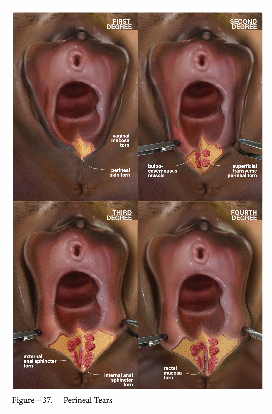
Comparison: Intact anatomy (left) versus first- and second-degree perineal injury (right). The two plates use identical anatomical framing and landmark labeling so the reader can map injury severity directly onto a known reference. Third- and fourth-degree injuries extend deeper into the anal sphincter complex and are addressed in subsequent plates.

Surgical Reference Plates

The remaining plates document procedural anatomy for operative delivery and surgical repair, completing the arc from diagnostic imaging through intervention. These were drawn to accompany the surgical technique chapters of the textbook.

Surgical procedure plates
Figures
Surgical procedure plates — operative anatomy for delivery and repair, indexed to the surgical technique chapters

Context

These plates were commissioned as part of a comprehensive obstetrics textbook. The brief was straightforward: produce clear, anatomically precise illustrations that a medical student or midwife could study from and a practicing obstetrician could reference during clinical decision-making.

The project was a study in restraint. Obstetrics illustration can easily drift into decorative territory, but the clinical use case demanded the opposite: every line had to earn its place. The vulvar and perineal anatomy plate, for example, labels every structure but keeps the illustration clean enough that a first-year student can trace each landmark without confusion. The neonatal trauma classification compresses an entire clinical decision tree into one visual reference. That compression, distilling a complex judgment into a single plate a clinician can consult at a glance, was the hardest and most rewarding part of the commission.

Other work

Pulmonary Embolism

Pulmonary Embolism

Medical Illustration

Actinic Keratosis

Actinic Keratosis

Medical Illustration

Want to learn about the human body
from the inside?

steven.oppong@gmail.com