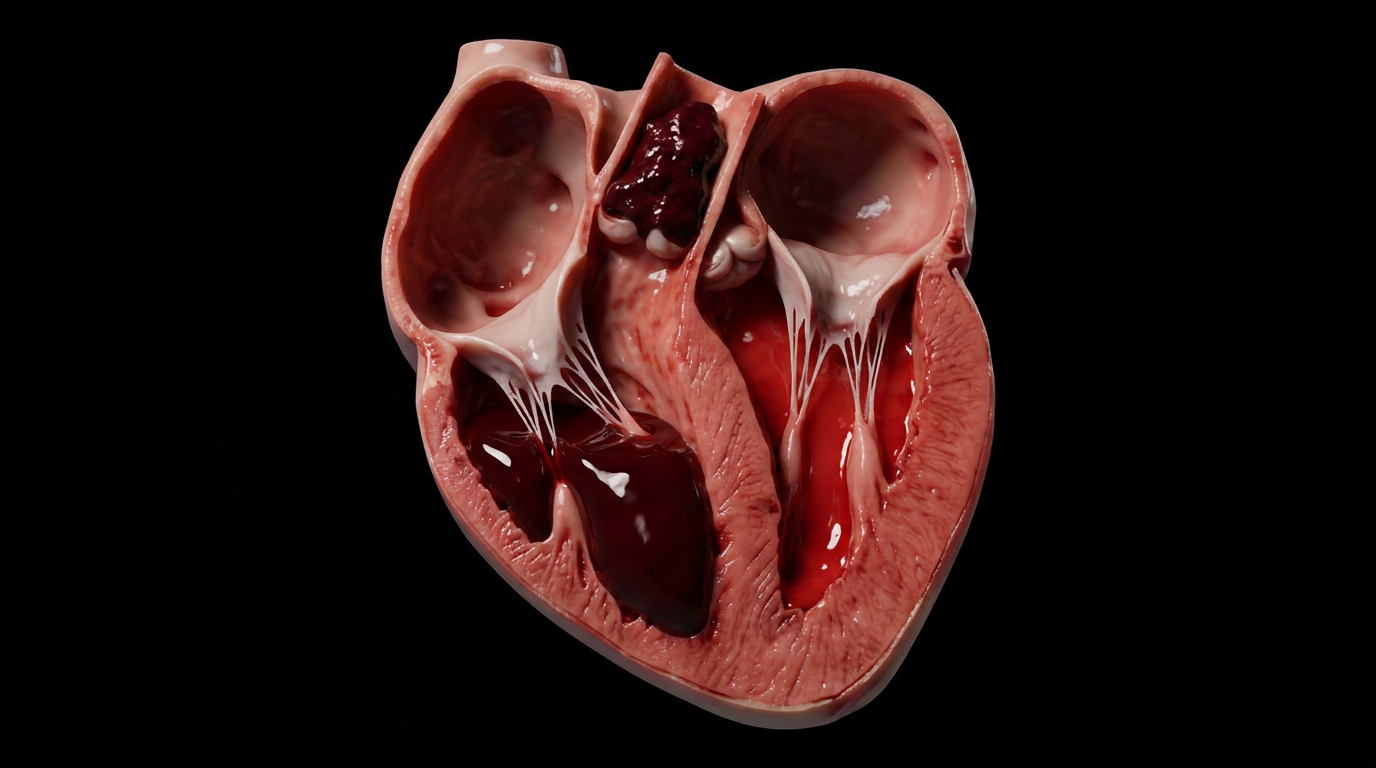
Sarcoidosis is a multisystem granulomatous disease of unknown cause. Somewhere, an antigen the immune system cannot identify or clear triggers a sustained Th1 response, and macrophages organize into tight, discrete nodules called granulomas. Unlike tuberculosis, the classic granulomatous disease it is most often compared against, these granulomas never develop central necrosis. Nothing dies at the center. The immune system simply keeps building, wall after wall, around a target it cannot name.
Almost any organ can be involved, but the lungs and their draining lymph nodes are affected in the overwhelming majority of cases. The disease's most recognizable imaging finding, bilateral hilar lymphadenopathy, is a direct consequence of granulomas accumulating exactly where lymphatic drainage from the lung converges.
Unlike tuberculosis, sarcoidosis has no identified causative organism. Whatever triggers it, real or environmental, is presented by macrophages and dendritic cells to naive CD4 T-cells, which polarize into a sustained Th1 response. Interferon-gamma and TNF-alpha drive continued macrophage activation and recruitment. Because the antigen is never cleared, the immune response never resolves the normal way. It simply keeps building.
Activated macrophages transform into epithelioid histiocytes, so named for their resemblance to epithelial cells, and pack together edge to edge into a compact, discrete nodule. In sarcoidosis this granuloma stays solid all the way through. There is no central caseous necrosis, no soft cheese-like core the way there is in a tuberculous tubercle. Only a sparse, thin cuff of lymphocytes rings the outer edge, which is why pathologists sometimes call it a "naked" granuloma. That absence of necrosis, tightly packed and uniform, is the single histologic feature that distinguishes it at a glance.
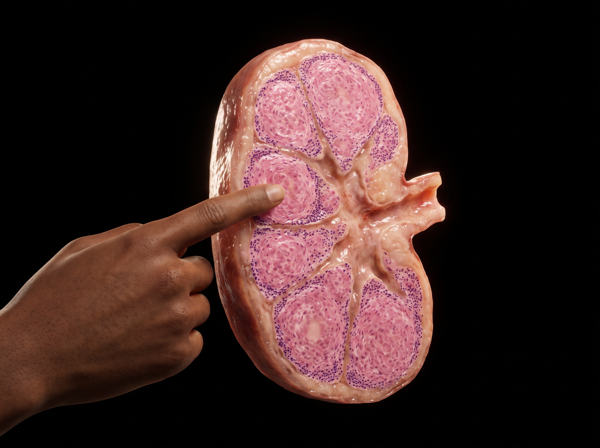
Sarcoidosis is systemic because macrophages are everywhere. The lungs and intrathoracic lymph nodes are involved in roughly nine out of ten cases, but granulomas can just as easily form in the skin, the eyes, the heart, the liver, or the nervous system. In the chest specifically, granulomas accumulate at the hilum, where lymphatic drainage from both lungs converges, producing the disease's signature finding on imaging: bilateral, symmetric hilar lymphadenopathy. It is often the first clue that brings a patient to a chest x-ray in the first place, sometimes before any other symptom appears.
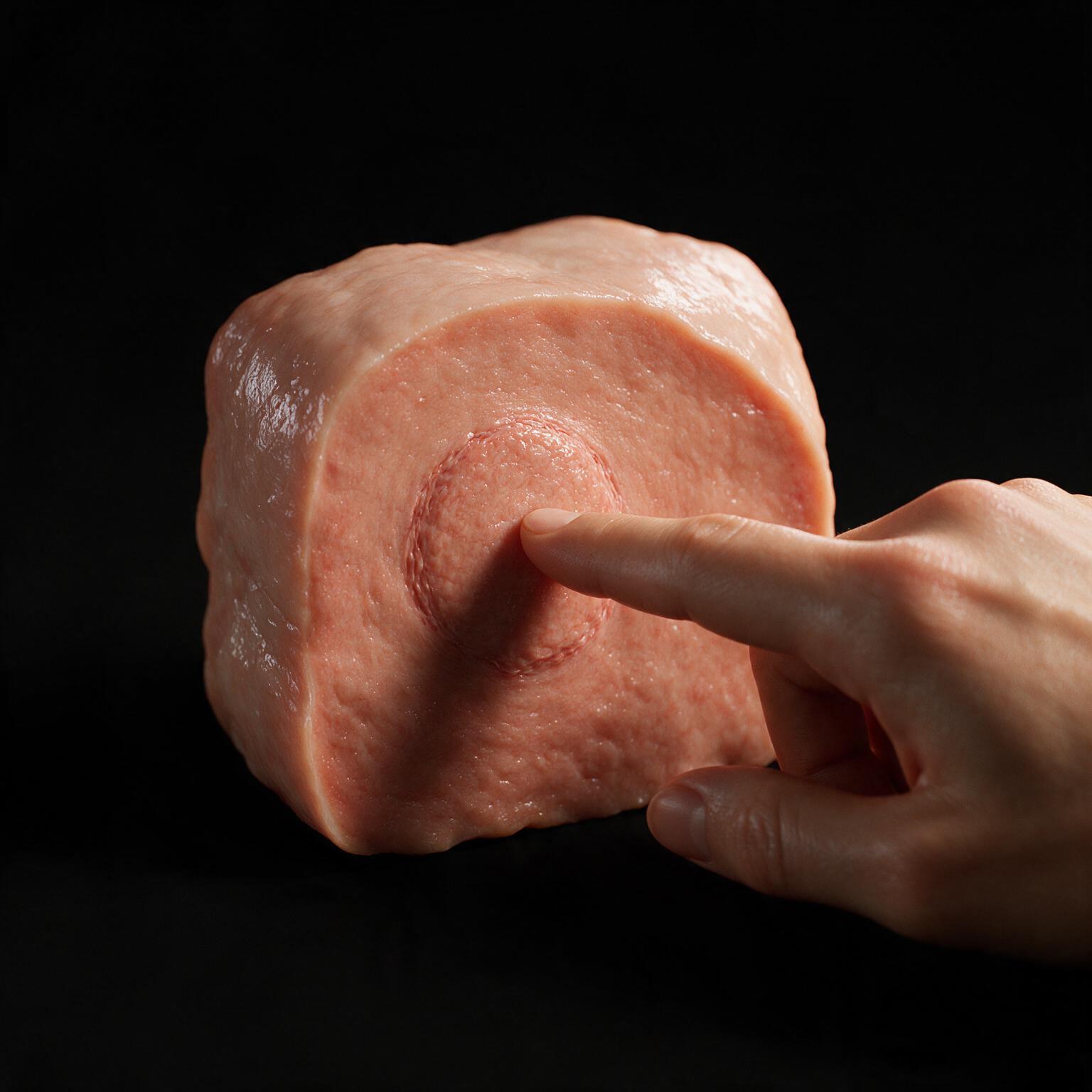
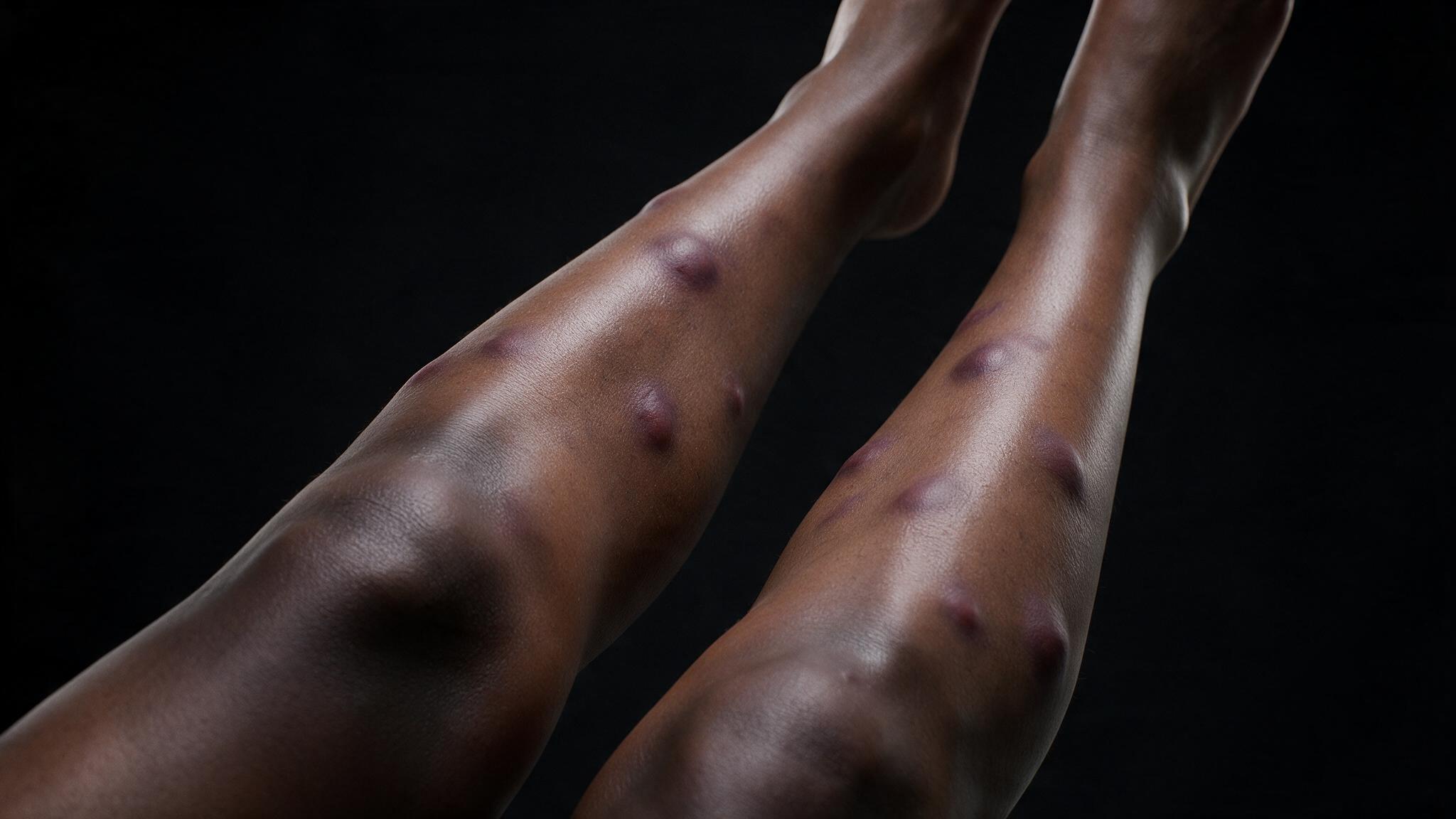
The same cross-reactive T-cells building granulomas in the lung do not always stay contained there. In a subset of patients they also attack the fibrous septa running between fat lobules in the subcutaneous tissue of the shin, which is exactly what produces the tender red nodules seen in the opening photograph. The septa become inflamed and packed with lymphocytes; the fat lobules themselves stay completely intact and necrosis-free. That septal pattern, inflammation strictly between the fat lobules rather than inside them, is what gives septal panniculitis its name.
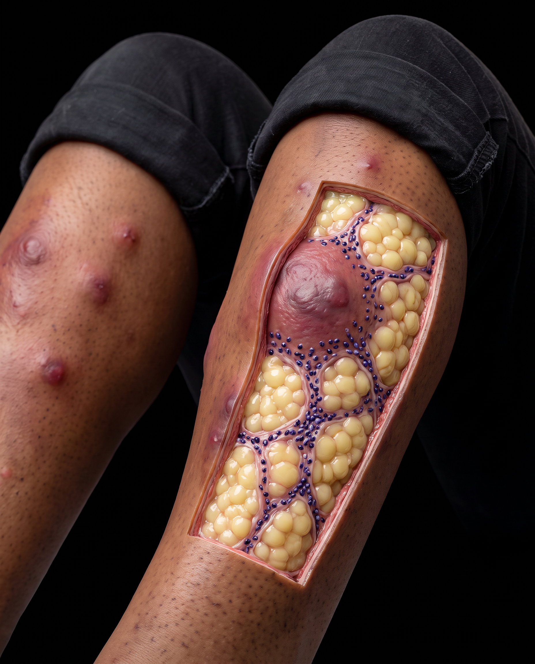
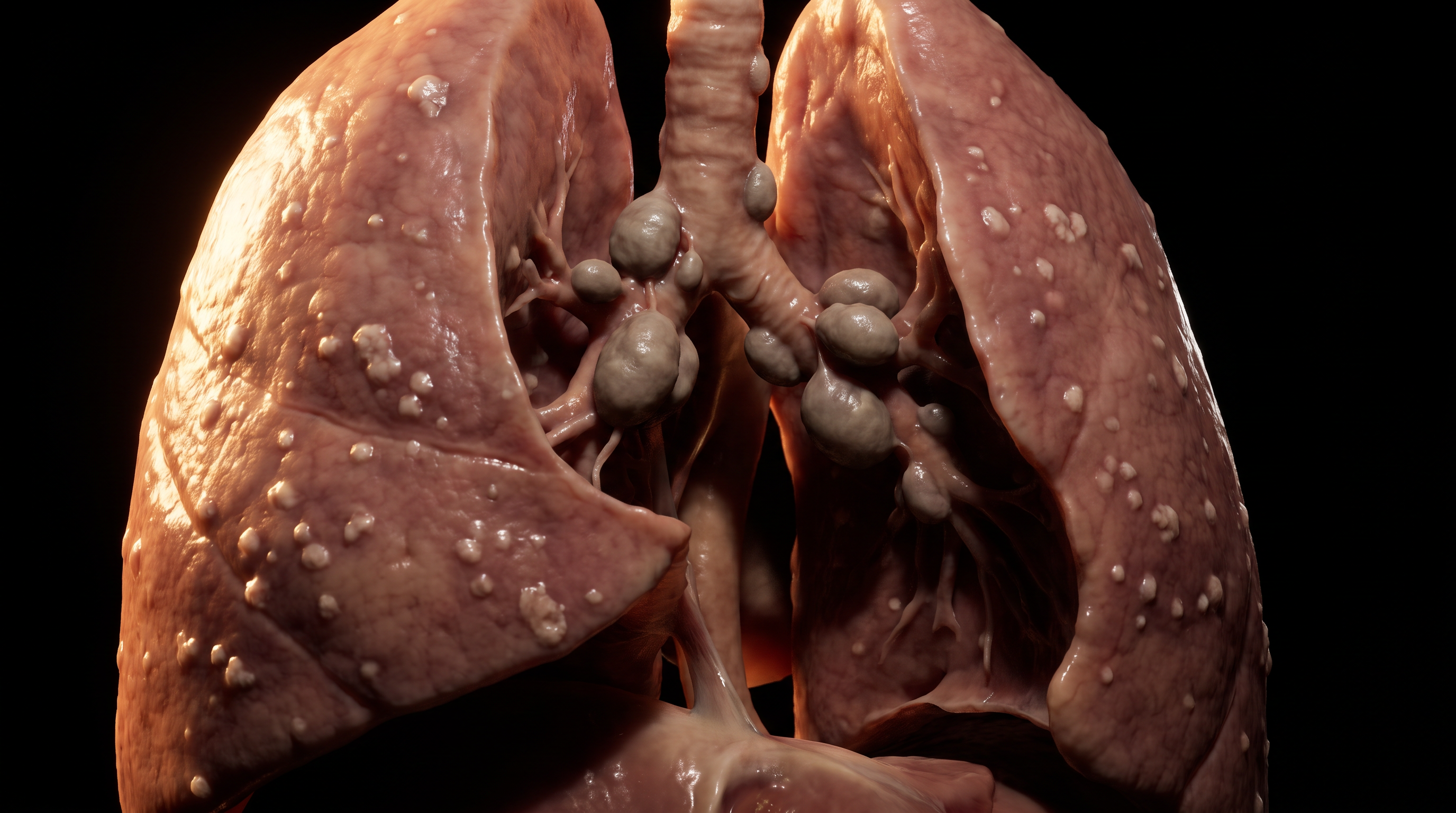
Sarcoidosis is a diagnosis of exclusion, confirmed histologically only once infectious and malignant causes of granulomatous disease have been ruled out. Course is genuinely unpredictable: a substantial proportion of patients undergo spontaneous remission within two years without ever needing treatment, while others progress to chronic pulmonary fibrosis or organ-threatening disease requiring long-term corticosteroids. One acute presentation carries a distinctly better prognosis than the rest. When bilateral hilar lymphadenopathy appears together with fever, joint pain, and tender red nodules on the shins, the constellation has its own name: Löfgren syndrome, the same shin nodules from the opening photograph, erythema nodosum, covered in detail in its own case study. Löfgren syndrome resolves on its own in the great majority of patients within months. The nodule on the shin, in this context, is not a warning of worse disease to come. It is often the sign that the body is already winning.
You're set. We'll send the full write-up to your inbox shortly.