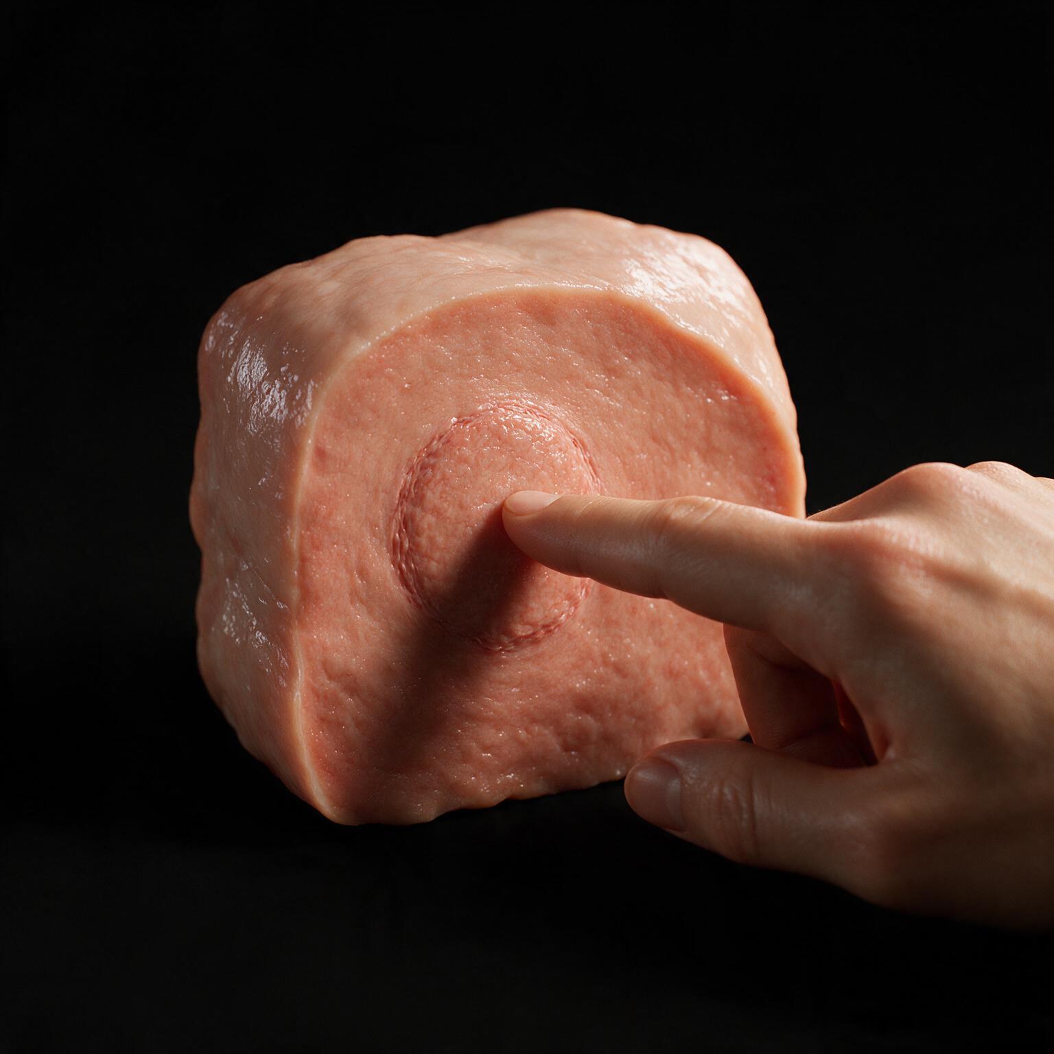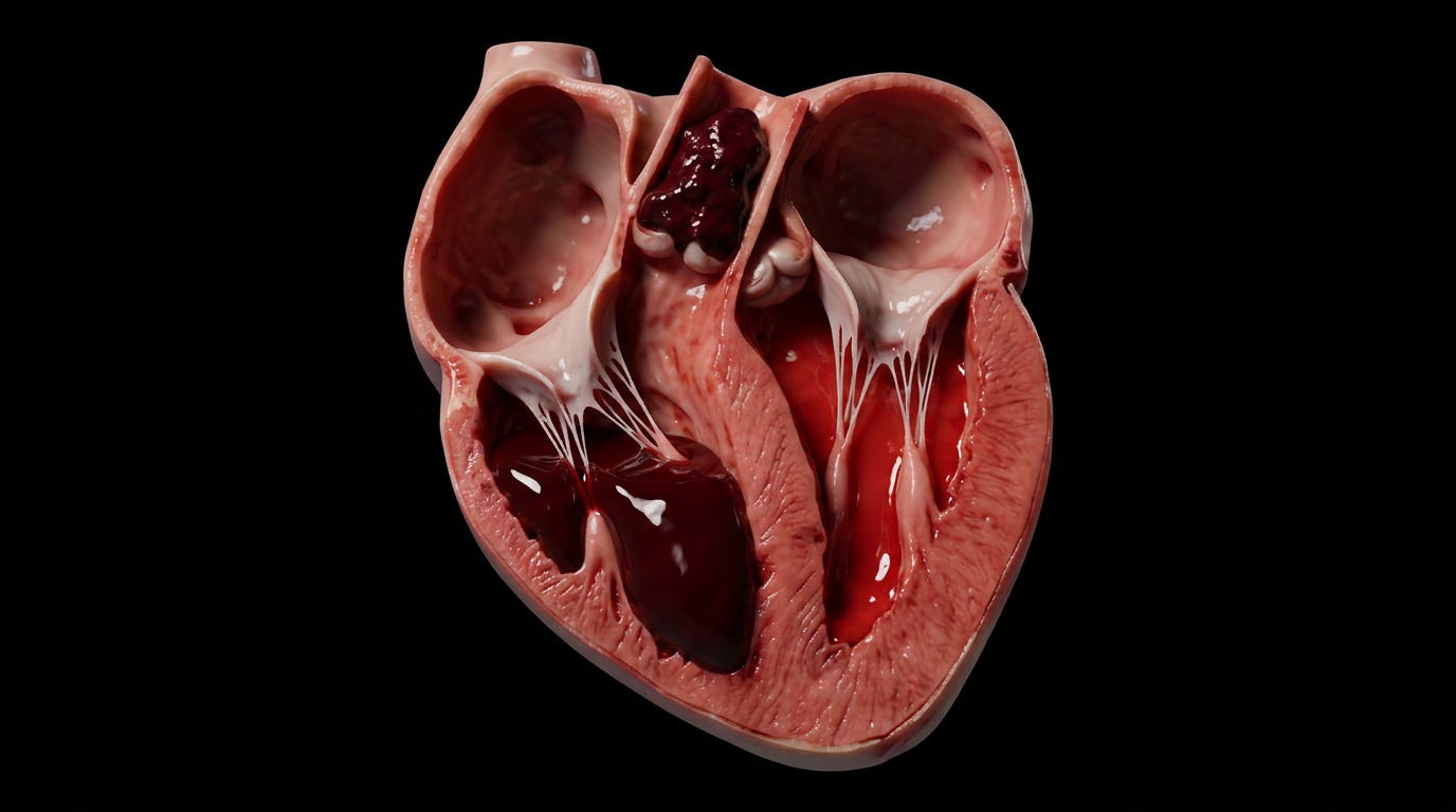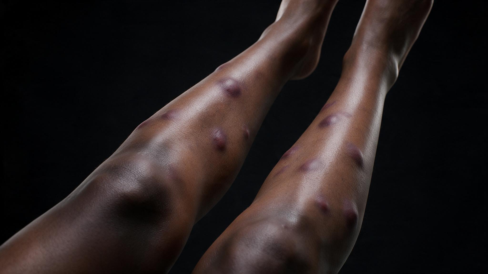
Erythema nodosum is a septal panniculitis. The inflammation lives in the fibrous septa that divide subcutaneous fat lobules, not in the fat cells themselves. It presents as tender, red nodules on the shins and is one of the body's most honest signals that something deeper is wrong, usually in the chest.
Two diseases converge on the same pulmonary node: tuberculosis and sarcoidosis. In both, macrophages in the lung sample an antigen and present it to T-cells. Those T-cells organize into granulomas, caseating in TB and non-caseating in sarcoidosis. That distinction matters for the chest, but it does not matter for the skin. The skin sees the same inflammatory consequence either way, because some of those primed T-cells also recognize the interlobular septa of subcutaneous fat as a secondary target.
In tuberculosis, alveolar macrophages engulf Mycobacterium tuberculosis. In sarcoidosis, an unknown antigen triggers the same cascade. Either way, the macrophage processes the antigen and presents it to naive T-cells in the lung-draining lymph nodes. Those T-cells activate, proliferate, and organize into granulomas. TB granulomas develop central caseous necrosis. Sarcoidosis granulomas remain non-caseating. Both achieve the same immunological outcome: a pool of primed T-cells now circulating through the body, looking for their target.
T-cell priming is not perfectly specific. The antigen that looks like mycobacterial protein or the unknown sarcoidosis trigger also resembles, just enough, an epitope expressed in the interlobular septa of subcutaneous fat. This is molecular mimicry. The T-cells leave the chest, circulate through the dermal and subcutaneous vasculature, and when they encounter the septal tissue that cross-reacts with their receptor, they mount a type IV hypersensitivity response. The septa become the collateral target of an immune fight that began in the lungs.
Within hours to days, neutrophils predominate in the interlobular septa, though early lesions already show a mixed infiltrate of lymphocytes and histiocytes alongside them. The septal walls thicken visibly. Small vessels within them dilate and leak. On histology, the fat lobules themselves remain pristine and untouched, their adipocytes clean and translucent. This is the defining feature of septal panniculitis, and it distinguishes erythema nodosum from lobular panniculitis, where the fat cells themselves are inflamed. The contrast between the clean fat lobules and the heavily inflamed septa surrounding them is the central diagnostic finding.
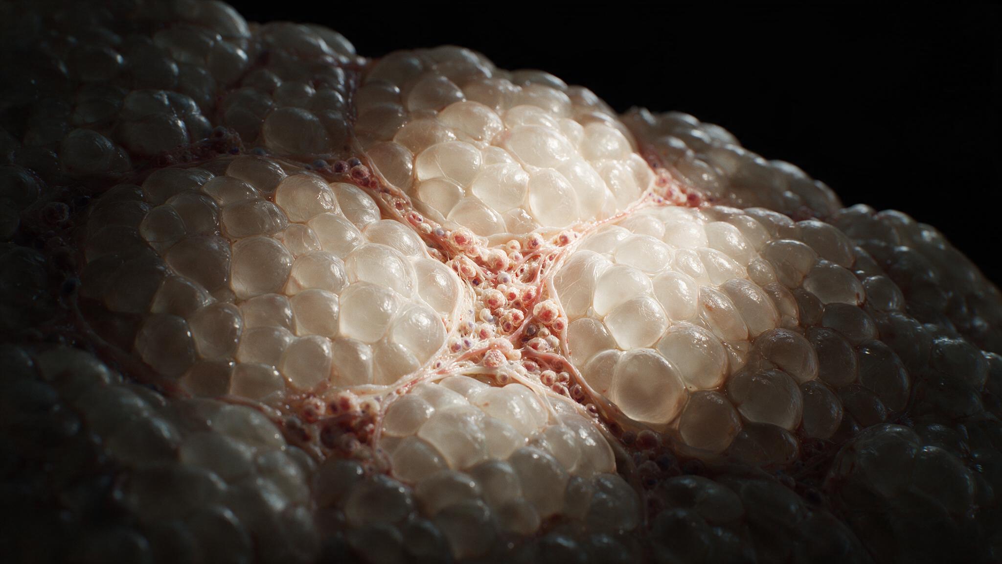
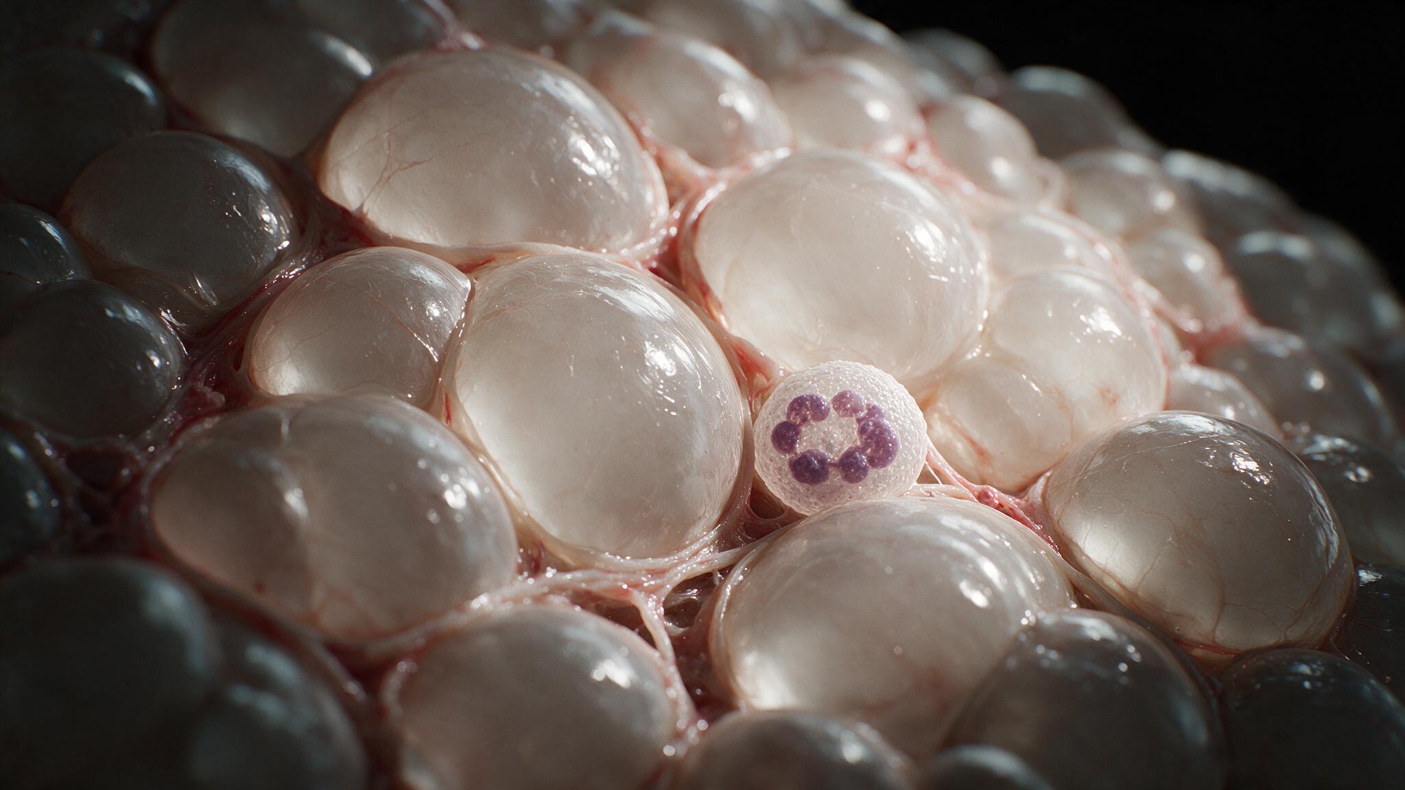
Over the following days to weeks, the cellular composition of the infiltrate shifts. T-cells and histiocytes come to dominate, while the early neutrophils recede. The septa now show a mixed chronic inflammatory infiltrate with lymphocytes as the majority population. Granulomatous inflammation can develop within the septa. Miescher's radial granulomas, small nodular aggregates of histiocytes arranged around a central cleft, are characteristic of erythema nodosum on biopsy. The neutrophils did the visible damage in the first wave. The lymphocytes arrived later and stayed, sustaining a low-grade inflammatory environment that can persist for weeks.
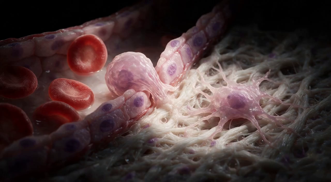
Erythema nodosum is not a primary skin disease. It is a delayed-type hypersensitivity reaction where the immune system, primed against a pulmonary pathogen or an unknown granulomatous trigger, has tagged the inter-lobular septa of subcutaneous fat as a secondary target. The macrophages started the conversation in the chest. The T-cells finished it in the skin. Treat the underlying driver, TB antibiotics or sarcoidosis management, and the nodules resolve. The fat was never the real enemy. It was collateral damage in a fight that began in the lungs. Understanding the two-wave infiltrate, neutrophils first, lymphocytes second, makes the timeline of clinical resolution legible. The red nodule on the shin is a chest x-ray waiting to be read.
You're set. We'll send the full write-up to your inbox shortly.