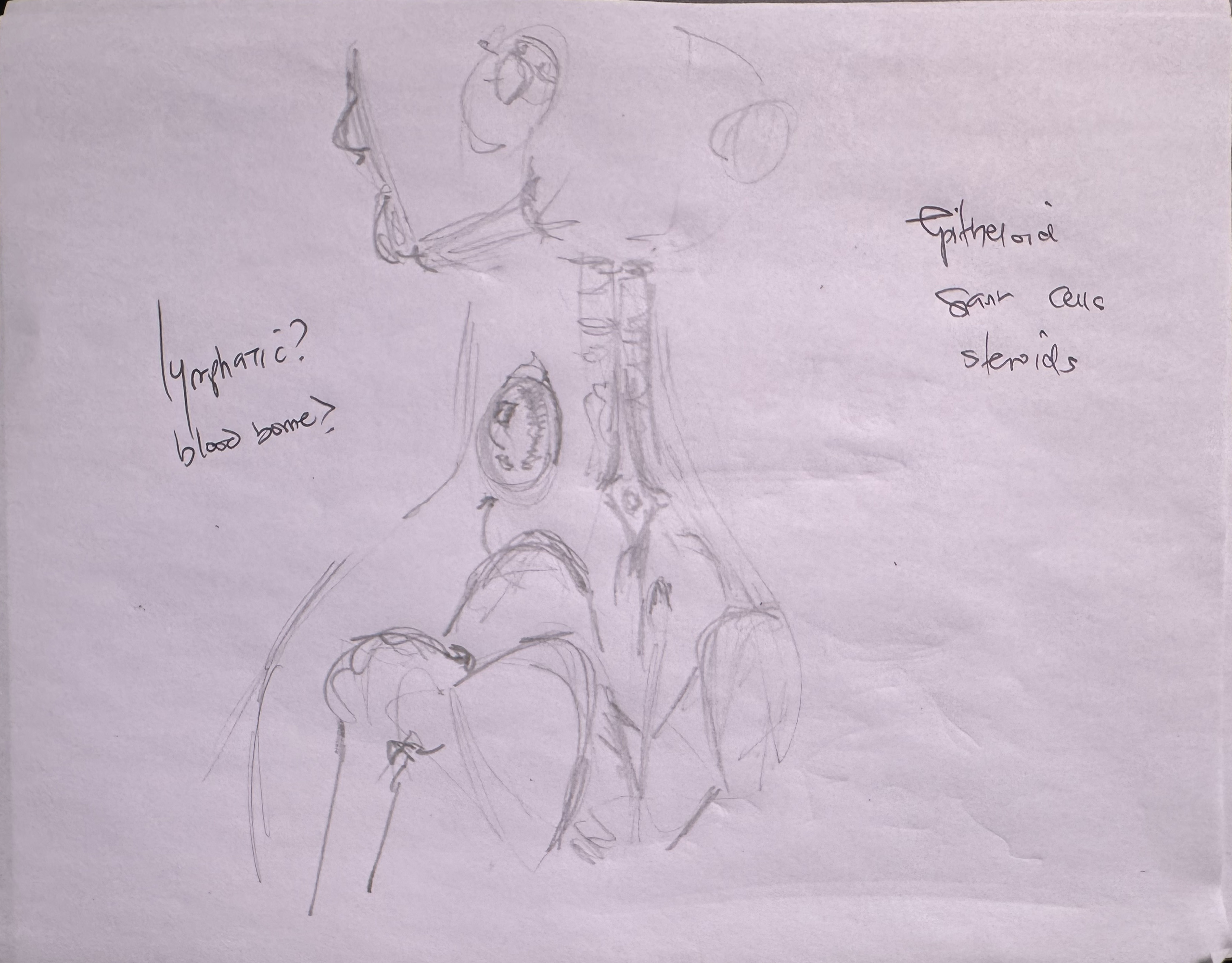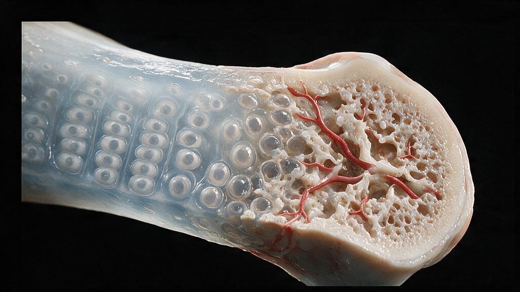
Bone is a ledger. Osteoblasts make deposits. Osteoclasts make withdrawals. Both sides must balance for the skeleton to stay strong, light, and properly shaped. In osteopetrosis, the withdrawal side fails: osteoclasts cannot resorb bone. The deposits continue. Bone mass accumulates, but it is brittle, disorganized, and architecturally incompetent. The marrow cavity fills in. Hematopoiesis is extinguished. Nerves passing through bony foramina are compressed. The bone that should be the body's scaffold becomes its prison.
Osteopetrosis is a family of rare genetic disorders caused by mutations in genes required for osteoclast function: carbonic anhydrase II, the proton pump ATP6i, the chloride channel ClC-7. Without a functional osteoclast, the resorption pit never forms. The ruffled border never develops. The acid never dissolves hydroxyapatite. And so bone, once laid down, stays. It accumulates in layers of woven bone that never remodel into organized lamellar bone. The X-ray is striking: dense, sclerotic bones with no corticomedullary distinction. The fragility is paradoxical: more bone, not less, but the bone is disorganized and brittle. Fractures are common. The only cure is hematopoietic stem cell transplant, which replaces the defective osteoclast precursors with functional ones.
Bone forms by two developmental routes. Endochondral ossification builds the long bones: a cartilage template forms first, then osteoblasts invade, mineralize the matrix, and replace cartilage with bone. This is how the femur, tibia, humerus, and vertebrae grow in length and shape. Intramembranous ossification skips the cartilage step: mesenchymal cells condense and differentiate directly into osteoblasts, laying down bone within a membrane. This is how the skull, the clavicles, and the flat bones of the face form. Both routes converge on the same endpoint: a collagen-apatite composite that is strong in compression and tension.
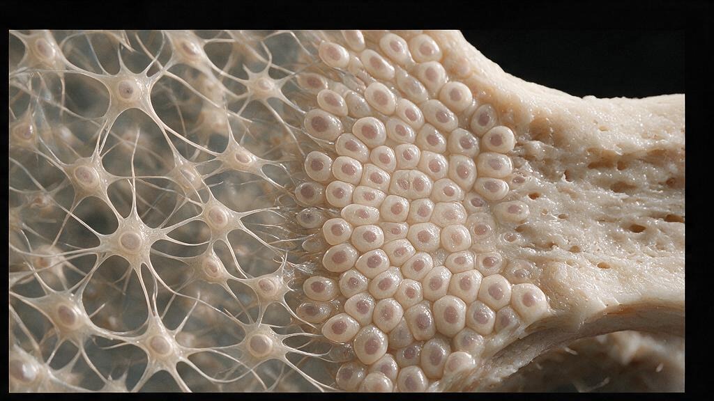
Newly formed bone is woven bone: collagen fibrils laid down in random orientations, osteocytes embedded haphazardly, mechanical properties isotropic and poor. Woven bone is temporary. Osteoclasts resorb it. Osteoblasts follow, laying down organized lamellar bone: parallel collagen sheets, osteocytes aligned along cement lines, mechanical properties anisotropic and optimized for load direction. This remodeling cycle, the basic multicellular unit, turns over the entire skeleton roughly every 10 years. In osteopetrosis, the osteoclast half of this cycle is absent. Woven bone accumulates. The remodeling never arrives. The result is a dense but fragile skeleton, like a building made of bricks with no mortar, stacked ever thicker but never reinforced.
The osteoclast is a polarized cell built for acid secretion. Its ruffled border, a folded plasma membrane, faces the bone surface and creates a sealed resorption compartment. Carbonic anhydrase II generates protons. The vacuolar H+-ATPase pump (ATP6i) pumps those protons across the ruffled border into the resorption lacuna. The ClC-7 chloride channel follows, maintaining electroneutrality by shunting chloride alongside the protons. The pH in the lacuna drops below 4.5. Hydroxyapatite dissolves. Collagen is exposed and degraded by cathepsin K. In osteopetrosis, any one of these molecular machines is broken. The acid cannot reach the bone. The mineral stays. The matrix stays. And the osteoclast, unable to do its job, sits on the bone surface like a locked door.
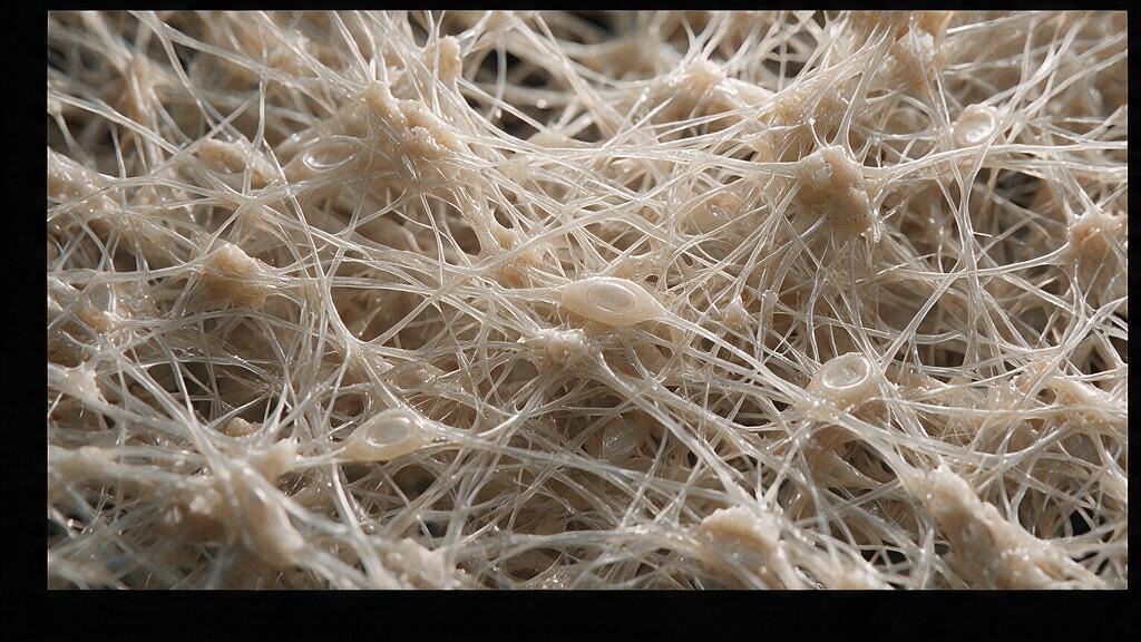
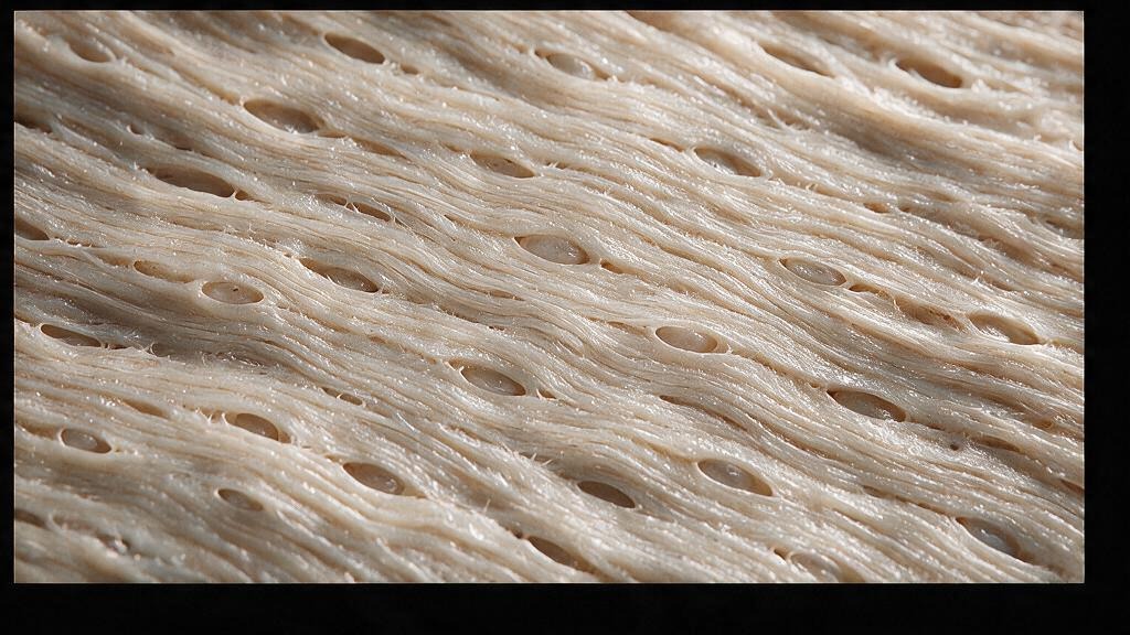
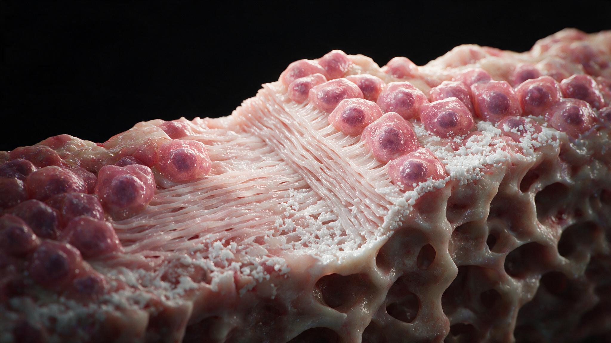
The infantile form of osteopetrosis is fatal within the first decade without treatment. Hematopoietic stem cell transplant from an HLA-matched donor provides functional osteoclast precursors. The donor-derived osteoclasts, now carrying intact CAII, ATP6i, and ClC-7, begin resorbing the accumulated bone. Marrow cavities reopen. Hematopoiesis resumes. Cranial nerve compression eases. But the transplant must come early, before the damage is irreversible. The adult form, Albers-Schonberg disease, is milder: patients live with dense bones, frequent fractures, and dental abscesses from compromised mandibular blood supply. There is no drug that fixes a broken proton pump. The illustrations in this series map the formation pathways and remodeling sequence that osteopetrosis arrests: endochondral and intramembranous ossification building the template, woven bone laid down as a placeholder, and lamellar bone as the final product that these patients never get to complete.
You're set. We'll send the full write-up to your inbox shortly.