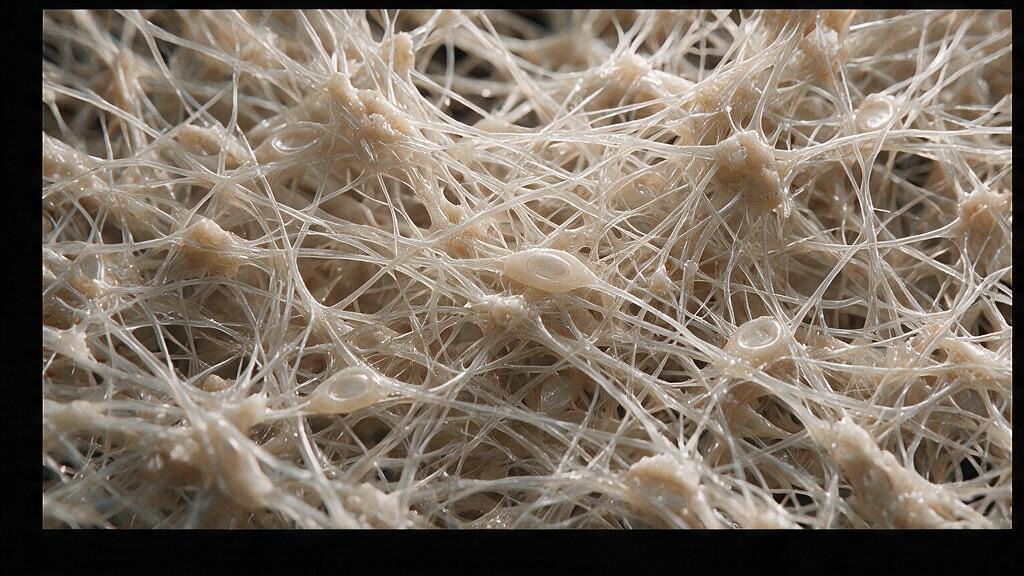
Bone is not a static mineral block. It is a living tissue that turns over constantly: osteoclasts dig resorption pits, osteoblasts lay down new osteoid, and that osteoid must mineralize, within days, into rigid hydroxyapatite. When mineralization fails, the osteoid seam stays soft. It accumulates. The bone is present, the collagen scaffold is there, but it never hardens. This is osteomalacia: soft bone, not from a lack of matrix but from a failure to mineralize it.
In children, the same defect produces rickets: growth plates thicken into unmineralized cartilage wedges, and weight-bearing bones bow under load. In adults, osteomalacia is subtler: diffuse bone pain, proximal muscle weakness, pseudofractures on x-ray. The cause is almost always vitamin D deficiency, calcium deficiency, or phosphate wasting. The treatment is replacement, not surgery.
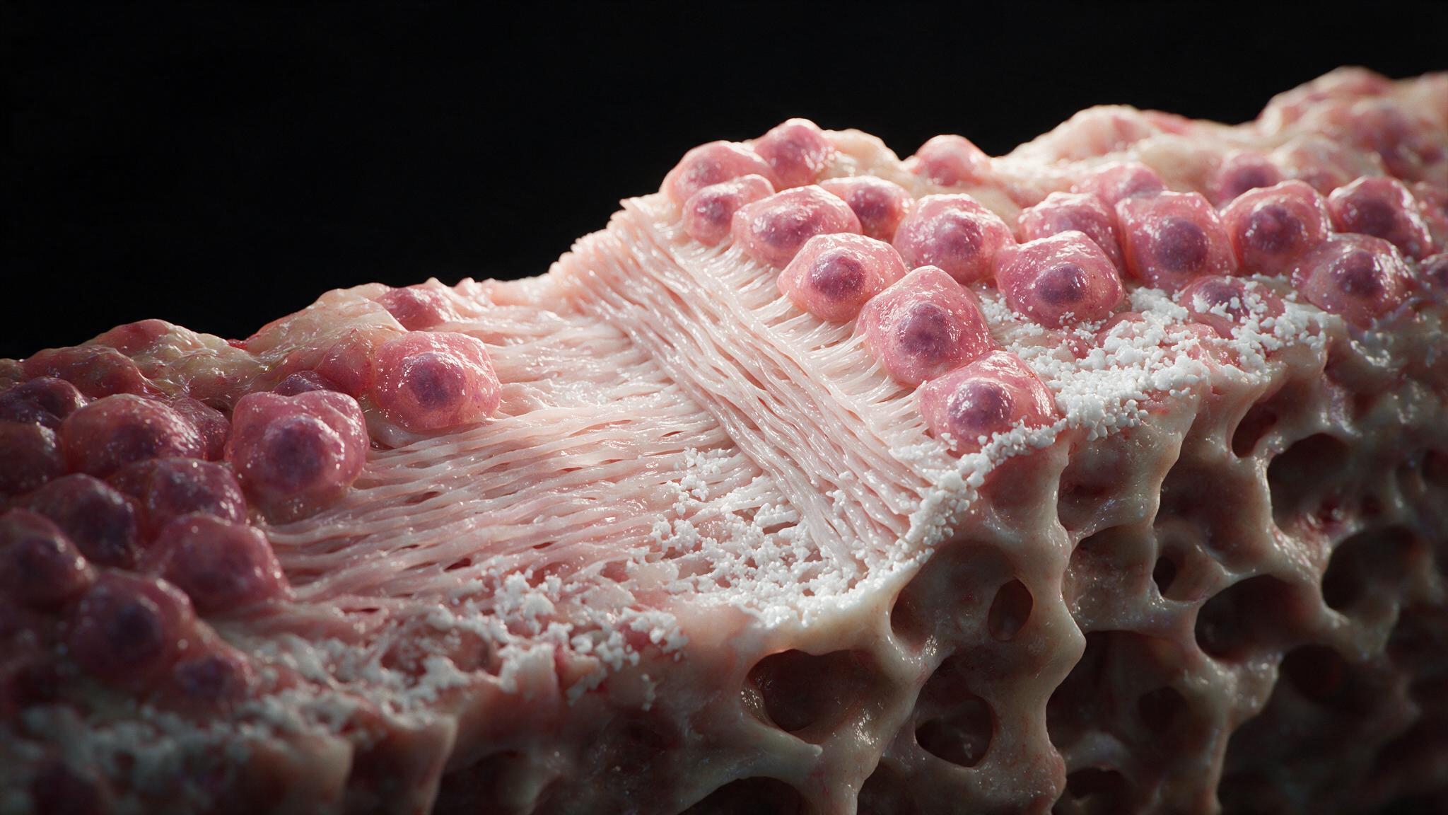
In healthy bone, osteoblasts secrete osteoid, a collagen-rich matrix, onto the bone surface. Within 10 to 15 days, this osteoid undergoes mineralization: calcium and phosphate precipitate as hydroxyapatite crystals along the collagen fibrils. The mineralization front is sharp. On a bone biopsy stained with toluidine blue, you see a thin osteoid seam (less than 5 lamellae thick) and a crisp transition from unmineralized matrix to calcified bone. Osteoblasts flatten into lining cells once their work is done. The process is rhythmic, ordered, and dependent on adequate calcium, phosphate, and activated vitamin D.
When vitamin D is deficient, calcium absorption from the gut drops. When phosphate is wasted, the substrate for hydroxyapatite formation disappears. In either case, the mineralization front stalls. Osteoblasts continue to lay down osteoid, but it never calcifies. The osteoid seam thickens: 10, 15, 20 lamellae deep, all unmineralized. Under the microscope, the seam stains pink and broad, with a ragged, patchy mineralization front. The bone surface looks busy with active osteoblasts, but they are producing matrix that will never harden. This is the histologic signature: increased osteoid volume, increased osteoid surface, decreased mineral apposition rate.
Unmineralized osteoid is mechanically incompetent. It deforms under load. The periosteum, richly innervated, stretches over softened bone and produces the dull, persistent ache that osteomalacia patients describe, often worse in the hips, low back, and thighs. Proximal muscle weakness follows: not from a primary myopathy but from pain-inhibited recruitment and disuse. On x-ray, Looser zones appear: narrow radiolucent lines perpendicular to the cortical margin, representing unhealed stress fractures filled with unmineralized osteoid. These pseudofractures are pathognomonic. The bone tries to repair itself, laying down matrix, but that matrix never calcifies. The cycle repeats until the deficiency is corrected.
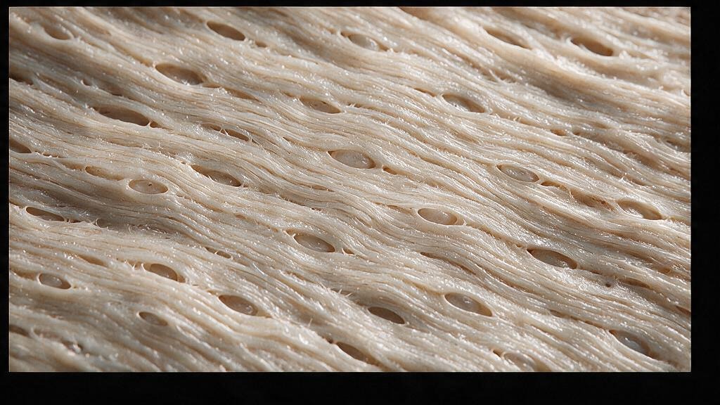
The treatment is replacement, and the response is measurable. Vitamin D3 (cholecalciferol) at 50,000 IU weekly for 8 to 12 weeks, followed by maintenance dosing, restores calcium absorption. The mineralization front sharpens within weeks. The osteoid seam thins as mineralization catches up to matrix deposition. Bone pain recedes. Pseudofractures heal. Muscle strength returns. The bone biopsy, if repeated, shows a normal osteoid seam width and a crisp mineralization front. What took months or years to develop can reverse in weeks once the substrate arrives. The illustrations in this series capture the histologic split: normal bone on one side, osteomalacia on the other, separated by the mineralization front that defines the difference between rigid and soft.
You're set. We'll send the full write-up to your inbox shortly.