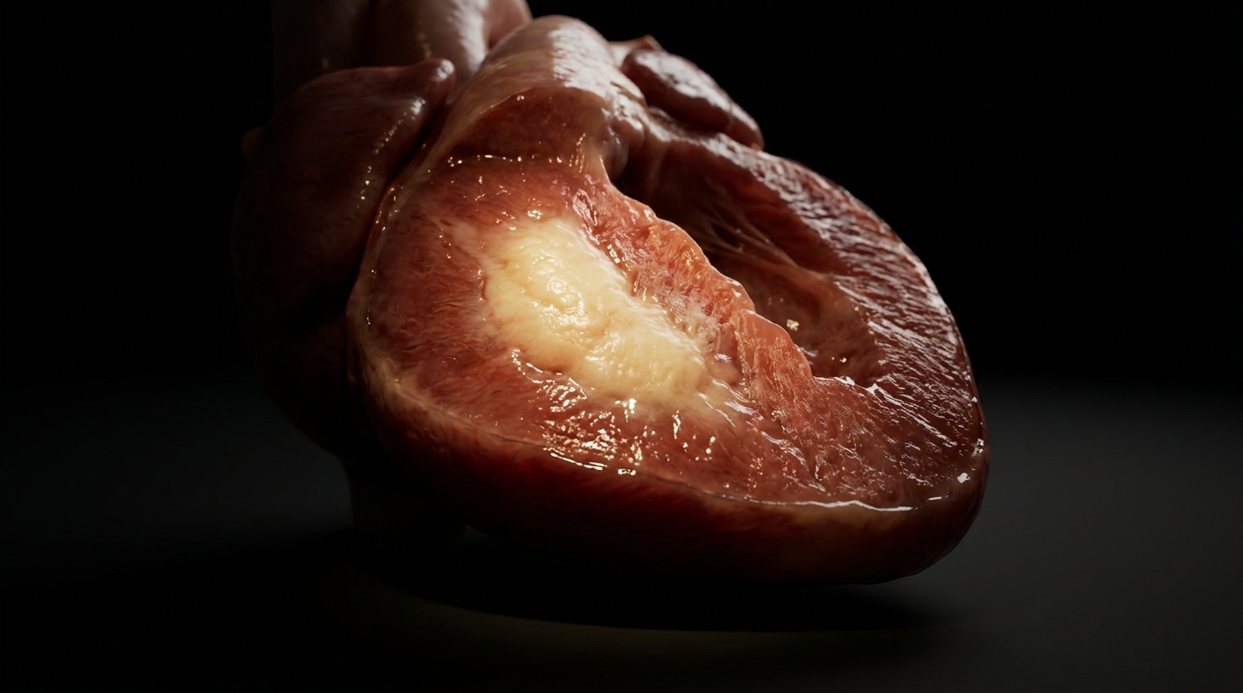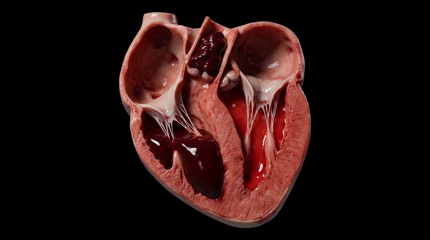
The liver receives a quarter of cardiac output. It has no choice but to absorb the pressure that the right heart cannot handle. When the right ventricle fails, that pressure backs up into the inferior vena cava, the hepatic veins, and finally into the hepatic sinusoids, distending the central veins and suffocating the centrilobular hepatocytes. The liver, caught between a failing pump upstream and its own metabolic demands, becomes congested, hypoxic, and eventually necrotic in a pattern as recognizable as a spice.
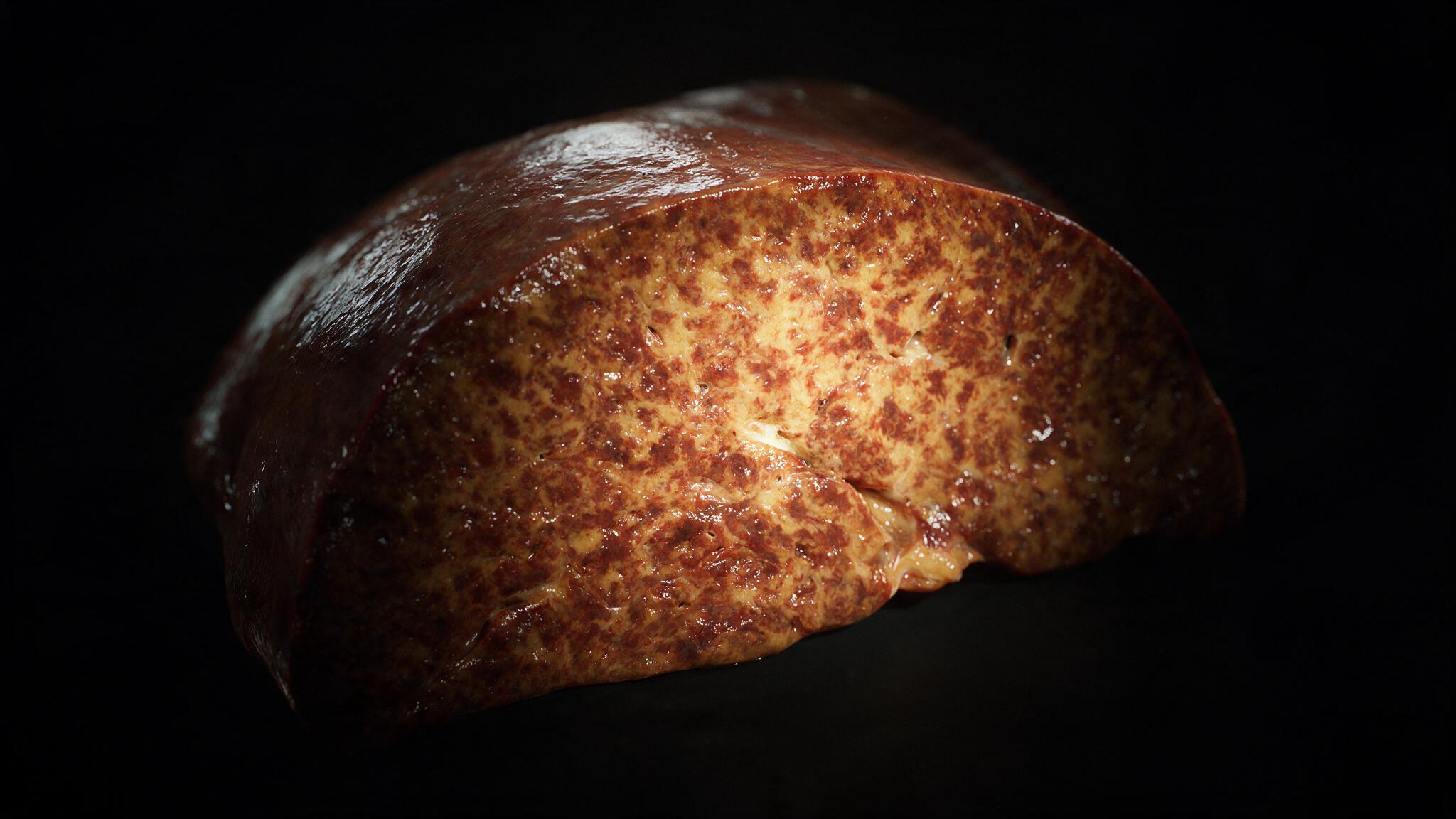
Cut into a chronically congested liver and the pattern is unmistakable: dark red-brown centrilobular zones alternating with paler periportal zones, a mottled appearance that pathologists have called nutmeg liver for over a century. It is the architectural signature of right-heart failure written in hepatic tissue. This case study maps the lobule from its normal architecture through congestion, necrosis, and the portal triad detail that makes the pattern legible.
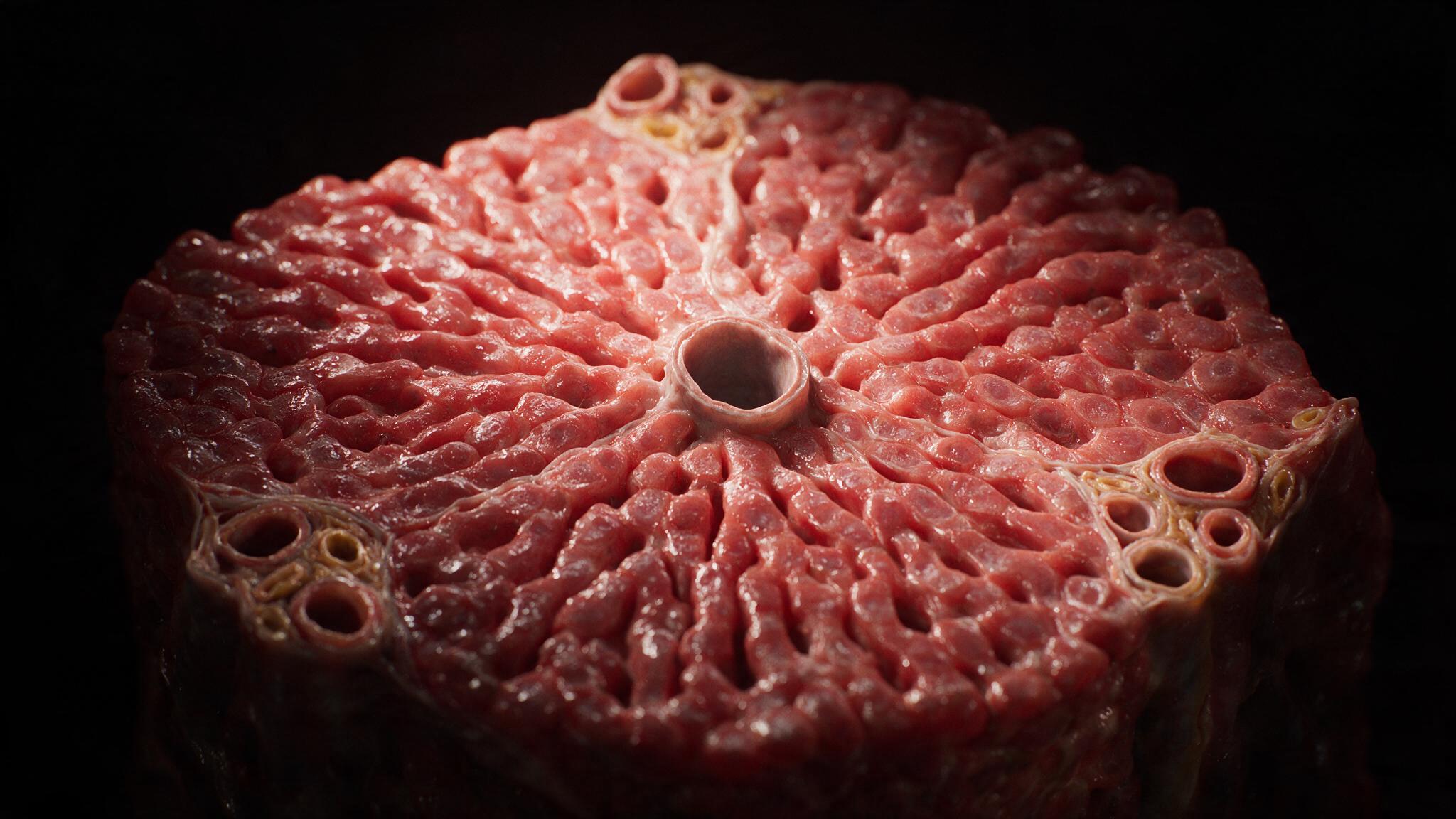
The hepatic lobule is organized around a simple hydraulic gradient. Blood enters from the portal triad at the periphery (portal vein and hepatic artery) and flows through the sinusoids toward the central vein, which drains into the hepatic vein and then the inferior vena cava. The sinusoids are low-pressure, low-resistance channels lined by fenestrated endothelium, designed to let plasma percolate freely into the space of Disse where hepatocytes can extract what they need. This design is efficient for metabolism but vulnerable to backpressure. When right-heart failure elevates central venous pressure, that pressure transmits directly through the hepatic veins into the central veins and sinusoids. The central vein, a thin endothelial tube with no muscular wall, distends. The sinusoids dilate. Blood pools. The lobule begins to drown from the center outward.
Hepatocytes are not equally vulnerable across the lobule. The periportal hepatocytes of zone 1 receive blood fresh from the hepatic artery, rich in oxygen and nutrients. The centrilobular hepatocytes of zone 3, at the distal end of the sinusoids, receive blood that has already passed through zones 1 and 2, its oxygen largely extracted. Under normal conditions, zone 3 operates on a thinner oxygen margin. Under congestion, that margin vanishes. The distended sinusoids slow flow. Stasis prolongs the transit time. The already hypoxic blood in zone 3 becomes frankly anoxic. Hepatocytes swell, their nuclei condense and fragment, and the centrilobular zone undergoes coagulative necrosis. The gross correlate is the nutmeg pattern: each dark spot on the cut surface is a congested, necrotic zone 3. Each pale ring around it is surviving zone 1 and 2 tissue, still perfused.
To understand why zone 3 dies first, you must understand what zone 1 has that zone 3 does not: proximity to the portal triad. Each triad bundles three vessels: a branch of the hepatic artery (thick muscular wall, small round lumen, bright oxygenated blood), a branch of the portal vein (thinner wall, larger irregular lumen, darker venous blood), and a bile duct (simple cuboidal epithelium, small lumen filled with golden viscous bile). Blood from the artery and vein mix in the sinusoids at the lobule periphery, creating the oxygen-rich inflow that zone 1 enjoys. Zone 3, at the opposite end of the lobule, gets what remains. The triad is the life support of the lobule. Its anatomy explains the gradient of injury. The congestion sequence in this series was built to make that gradient visible: the distended central vein, the pooled blood, the necrotic pericentral hepatocytes, and the surviving periportal tissue still anchored to its triad.
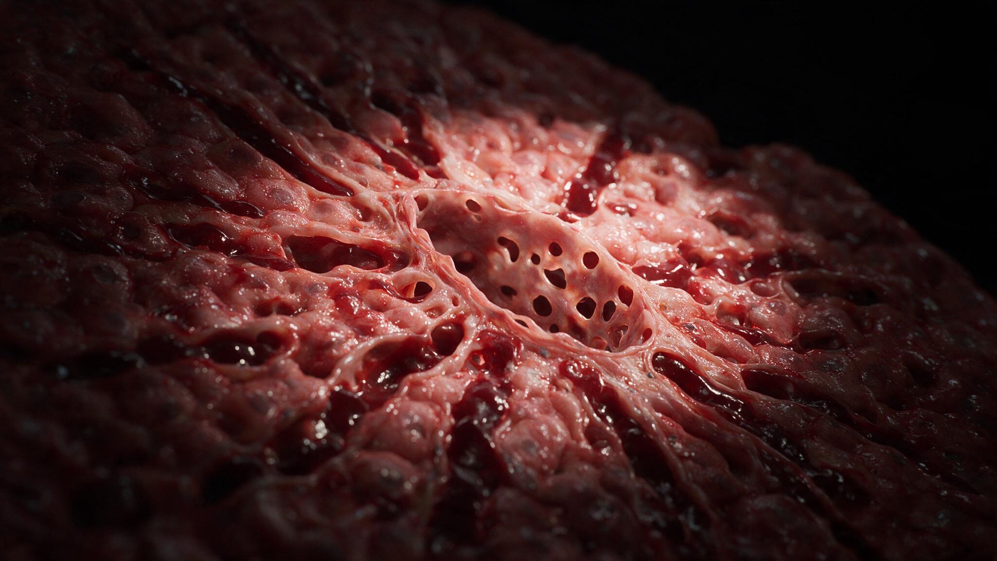
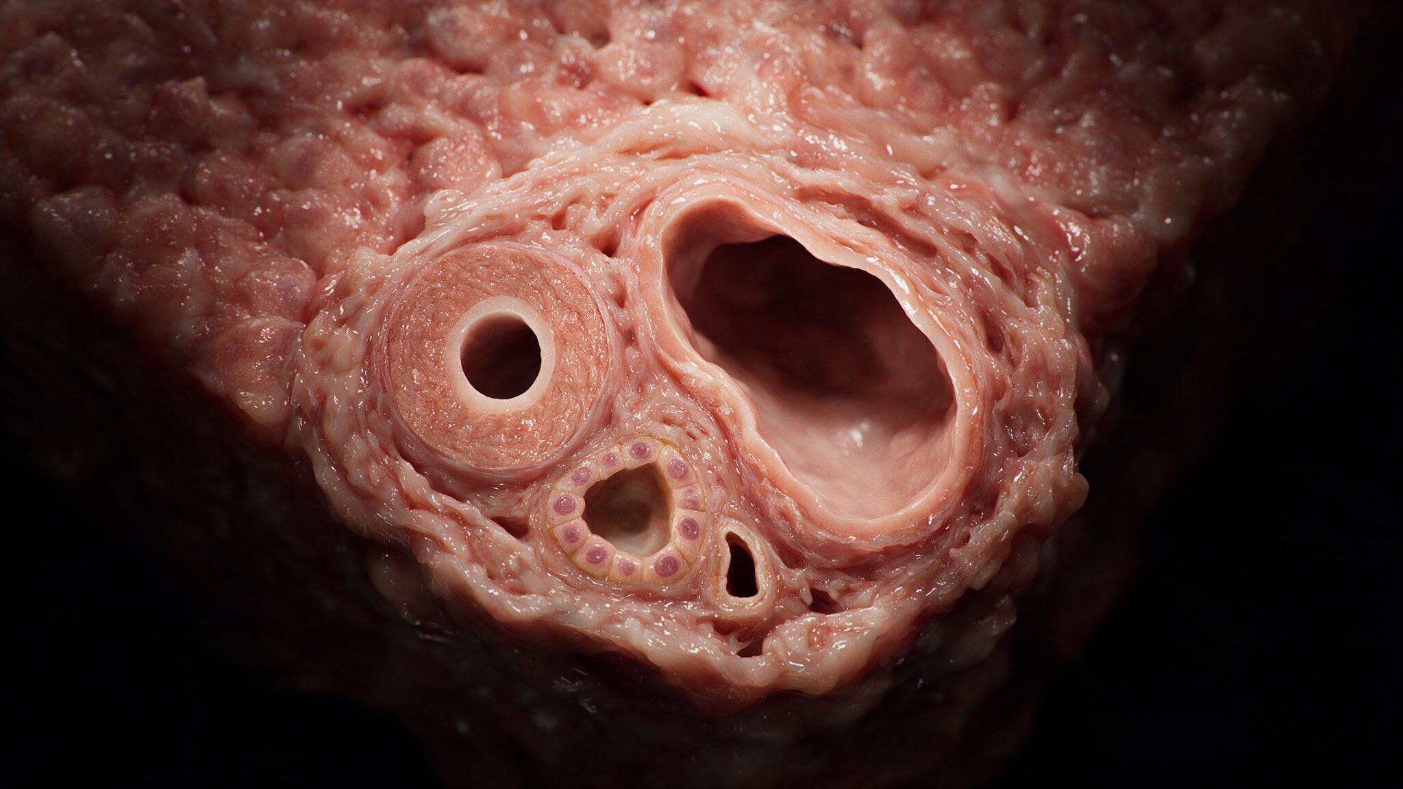
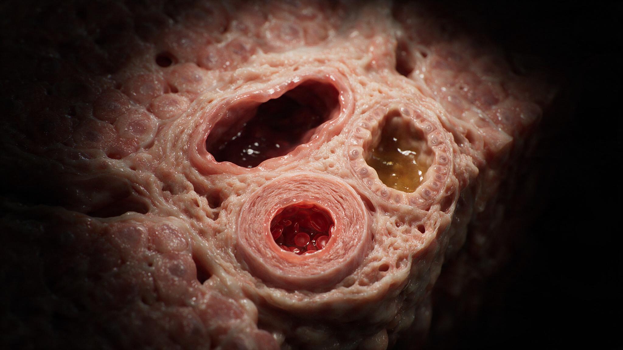
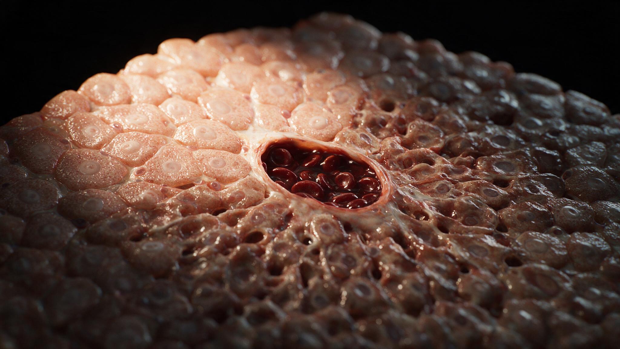
The nutmeg liver is a warning that the heart is failing. Treat the heart failure — diurese the fluid, reduce the afterload, restore forward flow — and the congestion recedes. The central vein pressure drops. The sinusoids drain. Zone 3 hepatocytes, if not yet necrotic, recover. But chronic, repeated episodes of congestion remodel the liver. Centrilobular fibrosis develops. Collagen bridges form between central veins. The architecture distorts toward cardiac cirrhosis, a condition in which the liver becomes fibrotic not because hepatocytes are injured by a toxin, but because they are chronically starved of oxygen by a failing pump upstream. The illustrations in this series follow the pressure gradient from the triad inflow to the central vein outflow, because the nutmeg pattern is a map of oxygen tension, and reading that map correctly tells you where the problem actually lives: not in the liver, but in the heart.
You're set. We'll send the full write-up to your inbox shortly.