When a coronary artery occludes, the myocardium downstream doesn't die right away. There's a window, roughly 20 to 40 minutes, where the myocytes are ischemic but still salvageable. Past that point, the damage can't be undone. The cell swells. The mitochondria rupture. Calcium floods in. Denatured proteins coagulate into a firm, eosinophilic mass that holds the ghost shape of the tissue it used to be.
That's coagulative necrosis, the dominant pattern of cell death in every solid organ except the brain. What sets it apart from liquefactive, caseous, fat, and fibrinoid necrosis is that the architecture survives, at least for a while. Cell outlines stay visible. The extracellular matrix holds. The dead tissue stays firm, not liquid, until neutrophils and macrophages show up days later to digest it. This case follows that arc, from the pale infarct you can see with your own eyes down to the tombstone myocyte that only shows up under a microscope.
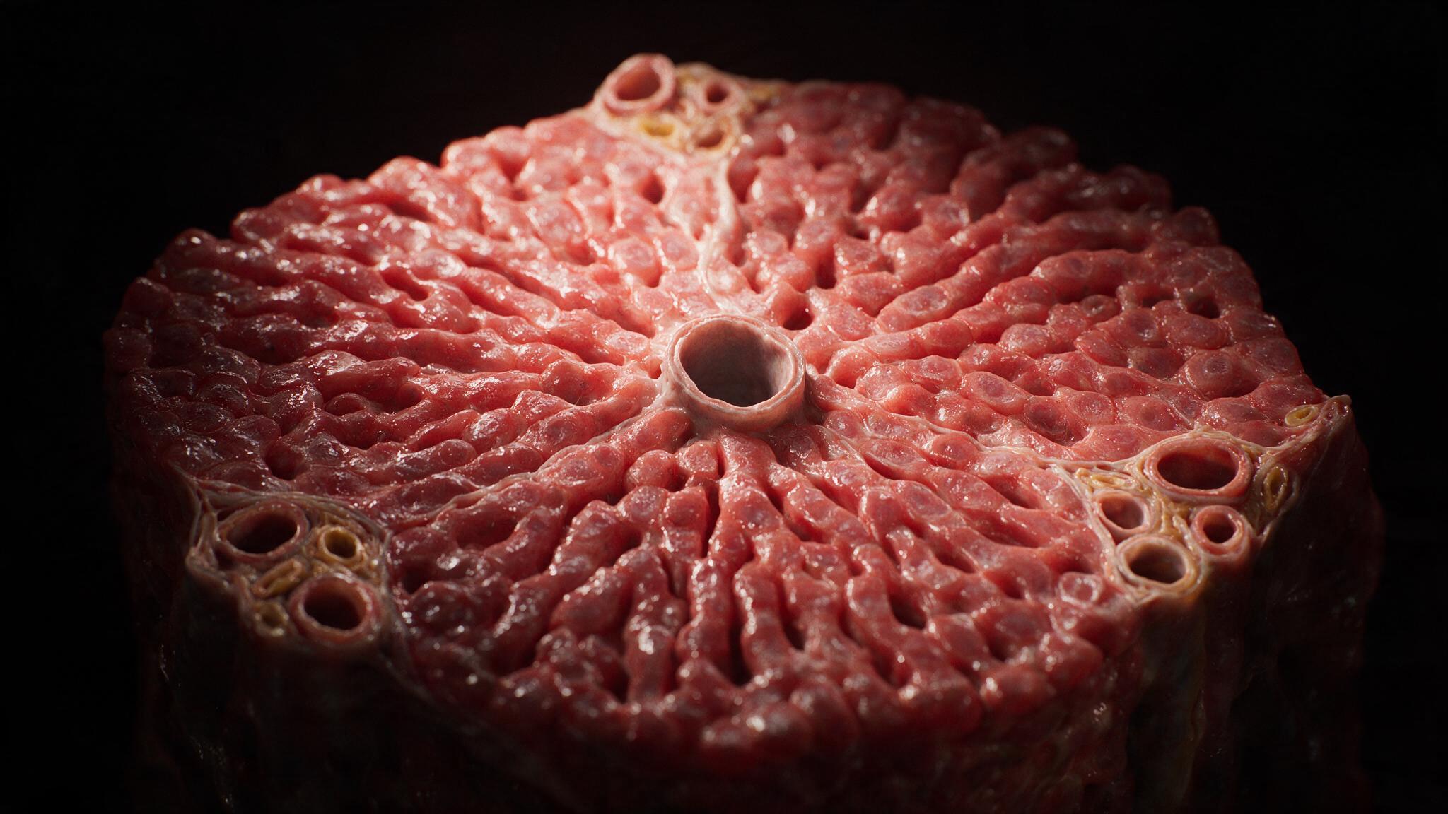
This story doesn't start with the infarct. It starts decades earlier, when LDL first slips into the arterial intima and oxidizes. That triggers an inflammatory response that pulls monocytes in. They turn into macrophages, gorge on the oxidized LDL, and become foam cells, the defining cell of the fatty streak, the earliest lesion you can actually see.
Over years, smooth muscle cells migrate in and build a fibrous cap over a growing lipid core. That's the mature plaque. And that cap is exactly what fails: it ruptures, exposes thrombogenic material to the blood, and triggers the clot that finally starves the muscle downstream. Everything that follows starts the moment that cap, decades in the making, gives way.
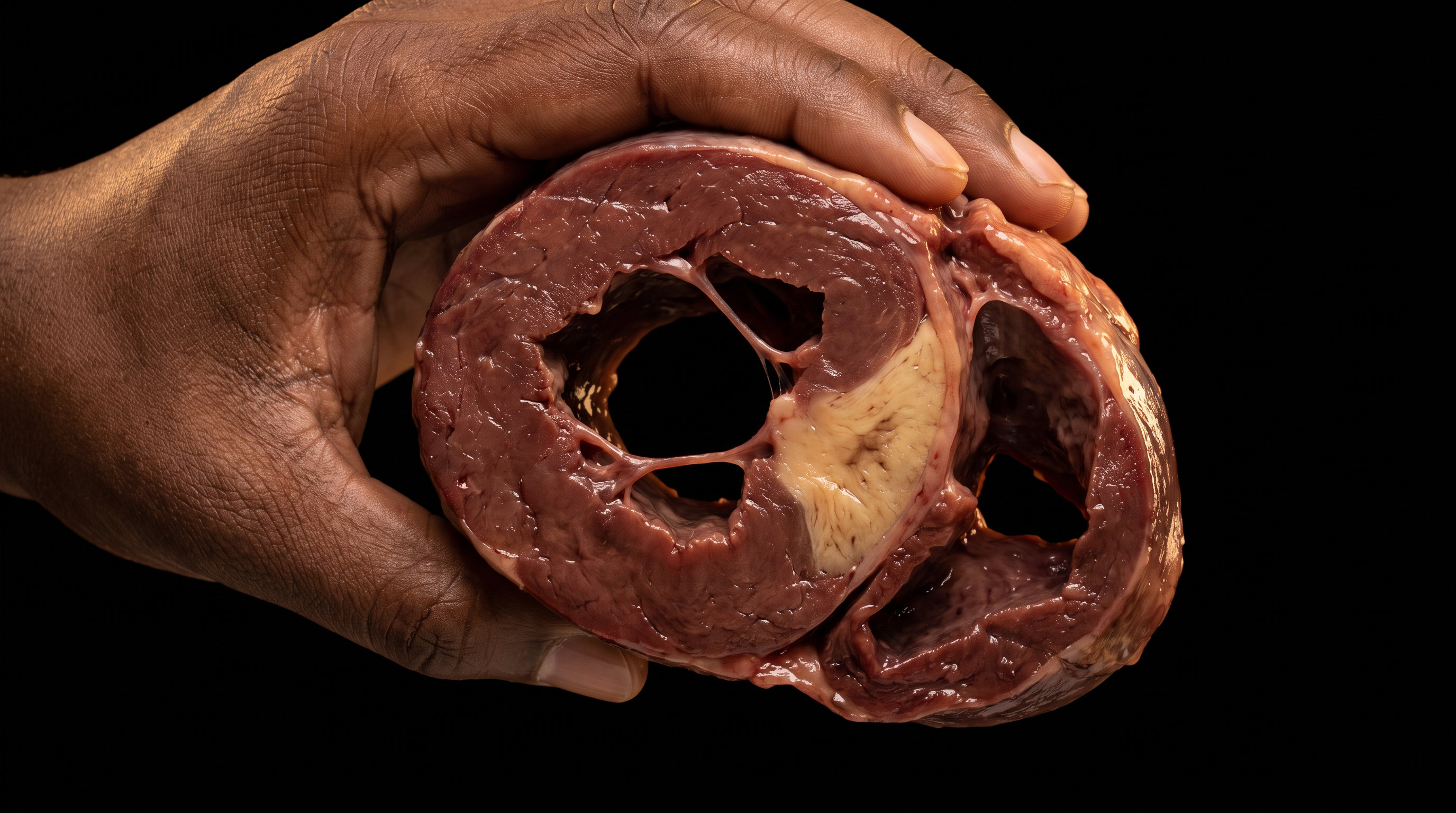
Twenty-four hours after the occlusion, you can see the damage with your own eyes. The infarcted zone turns pale, tan-yellow, slightly swollen, standing out hard against the dark red-brown of the muscle still alive around it. The border is sharp, often rimmed by a narrow band of hyperemia where vasodilation and inflammation are already underway.
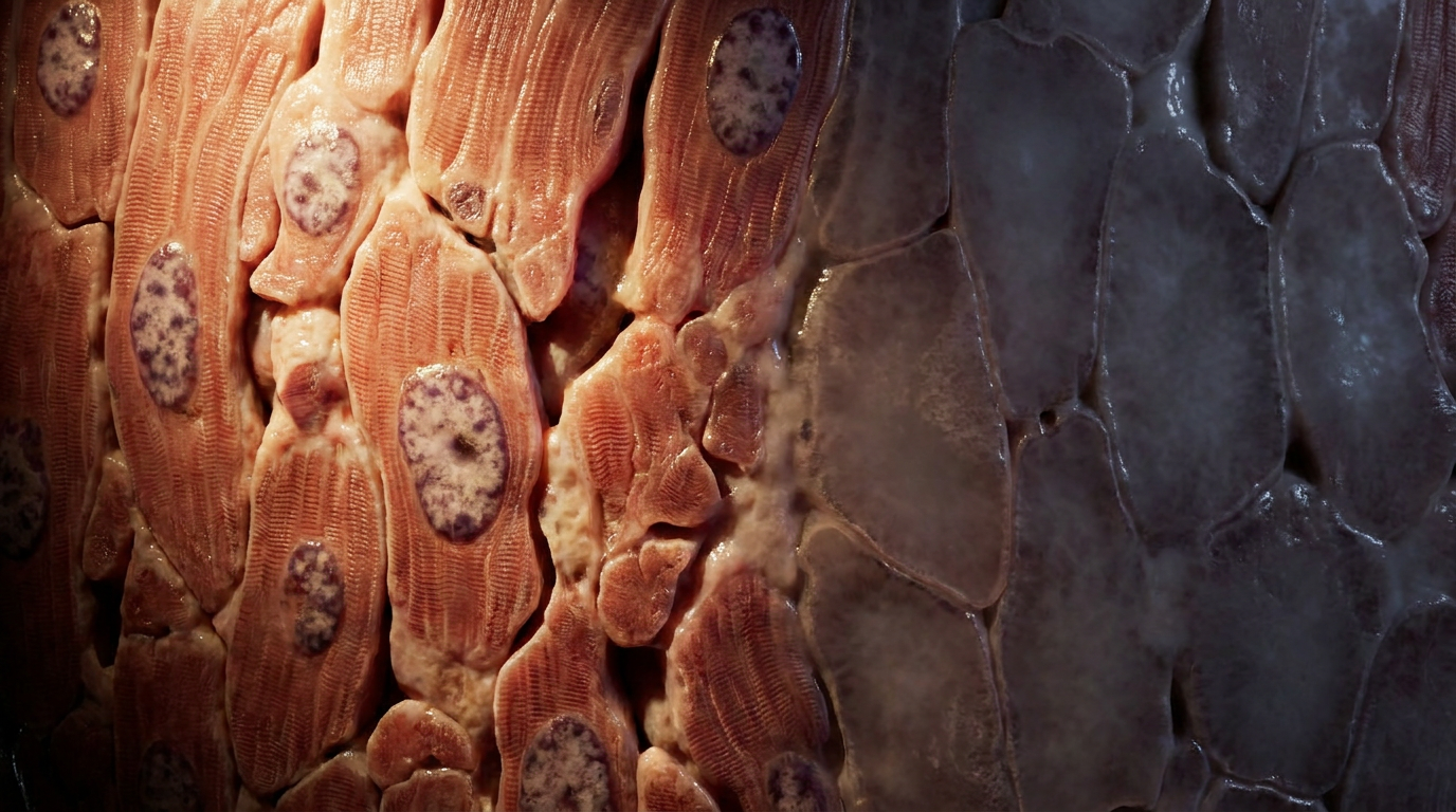
Touch it and it's firm, not soft, not liquefied, not friable. That firmness is the whole signature of coagulative necrosis: denatured structural proteins holding their shape as a solid mass. Under the microscope, the same story repeats at the cellular level. Hypereosinophilic myocytes have lost their nuclei but kept their outlines, a look so distinct pathologists just call them tombstone cells.
Under the microscope, the line between alive and dead is abrupt. Healthy myocytes show intact, elongated nuclei, dispersed chromatin, and clear cross-striations, the sarcomeric banding that actually drives contraction. Cross one narrow boundary and the story flips completely.
The nuclei are gone: pyknosis, karyorrhexis, karyolysis have erased them one by one. The cross-striations fade into a flat, glassy, deeply eosinophilic cytoplasm. But the sarcolemma, the cell outline, is still there, holding the shape of a cell that's already gone, like a cast of what used to live inside it. That's the tombstone: architecture intact, machinery dissolved. Even the extracellular matrix survives, collagen and elastin still in place, waiting to become the scaffold granulation tissue builds on later.
Which pattern a dead tissue follows comes down to one thing: how many lysosomes it had to begin with. Solid organs, heart, kidney, liver, spleen, are relatively poor in lysosomal enzymes. When their cells die, there's not enough enzyme around to digest the tissue quickly. The denatured proteins just hold their shape. The architecture stands. The dead zone stays firm.
Compare that to the brain, rich in lysosomes, where necrosis goes liquefactive: the tissue actually dissolves into a fluid-filled cavity within days. Or to a tuberculous granuloma, where necrosis goes caseous, a dry, cheese-like, friable mix of fragmented cells and lipid debris that doesn't resemble living tissue at all. Coagulative necrosis is the default for solid organs for a simple reason: their cells are built to work, not to digest themselves.
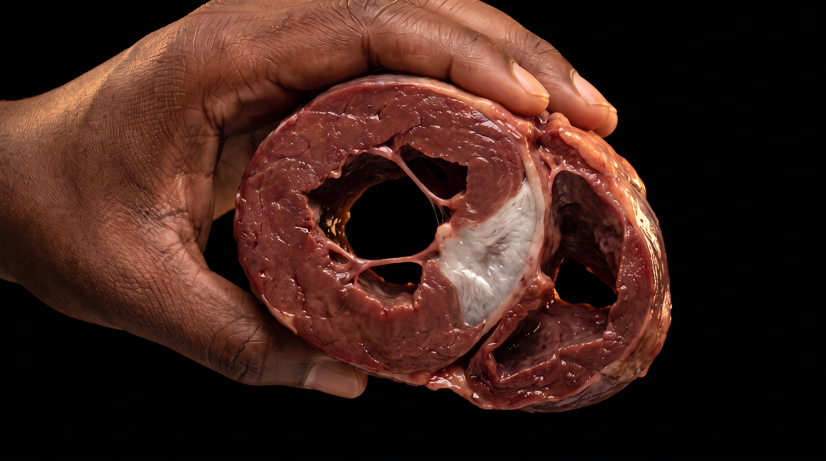
The infarct heals from the outside in. Neutrophils show up within hours, releasing enzymes that start the slow work of digesting the dead myocytes. Macrophages follow, clearing debris and calling in fibroblasts. Granulation tissue fills the gap, and over weeks to months collagen gets laid down until the infarct becomes a fibrous scar: permanent, non-contractile, a patch where working muscle used to be. The tombstone myocytes are gone by then, replaced by dense collagen, and the heart's architecture is changed for good. That's the whole arc this case traces: the gross scale, where a pale infarct stands out against muscle that's still alive, and the cellular scale, where a tombstone myocyte holds the outline of what it used to be while everything inside it dissolves.
You're set. We'll send the full write-up to your inbox shortly.