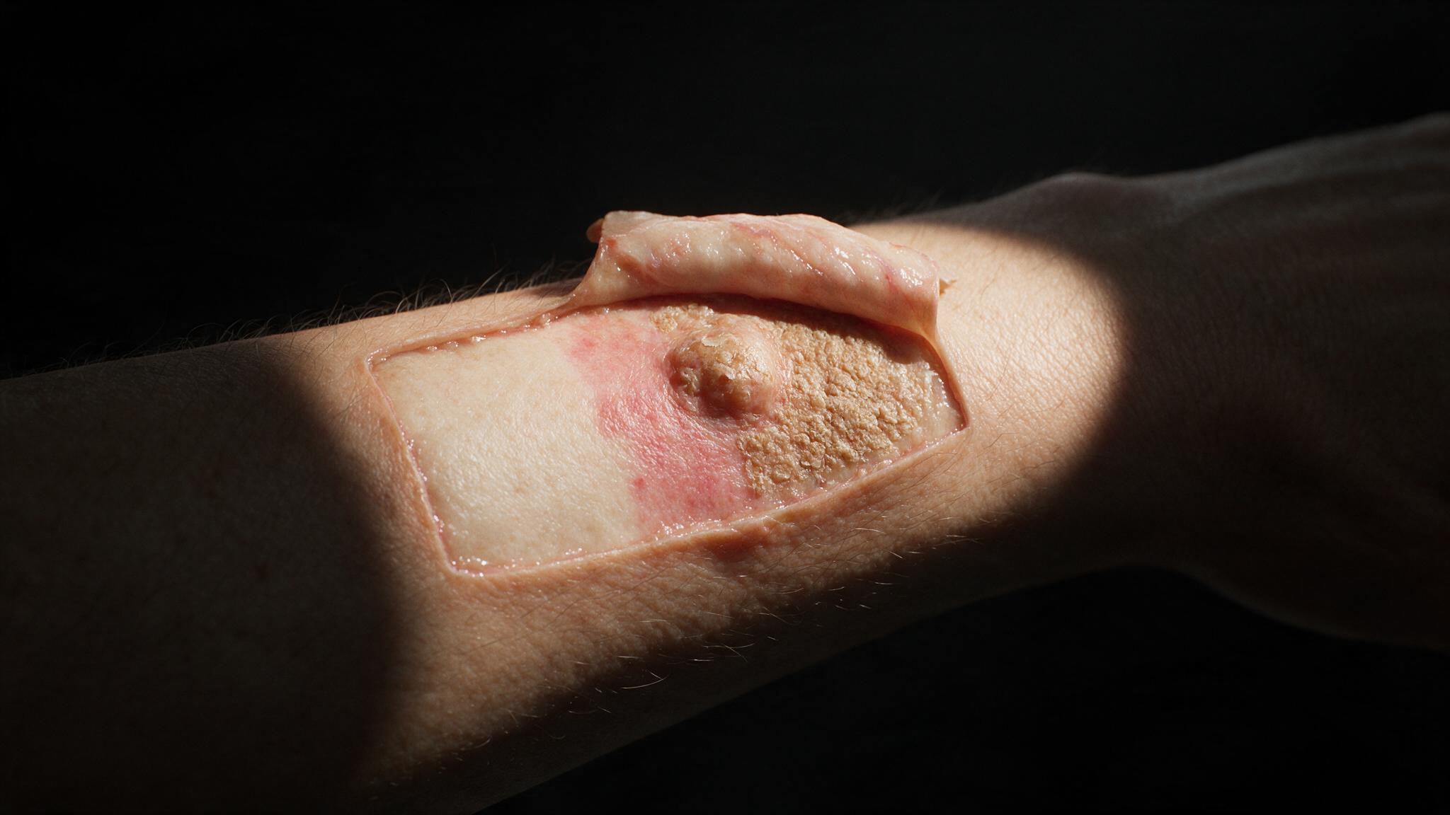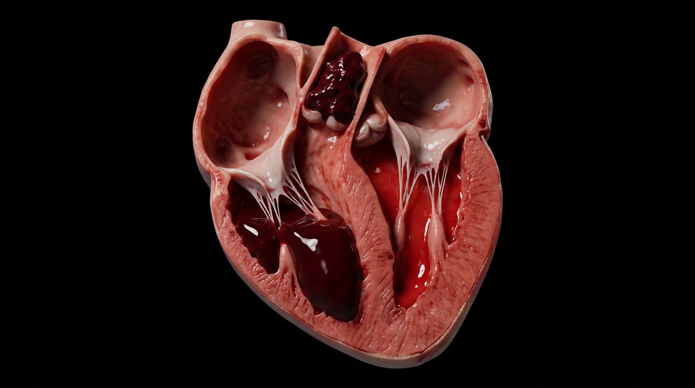
Portal hypertension is a pressure problem that becomes a tissue problem. When the liver stiffens, blood backs up through the portal venous system, elevating pressure throughout the splanchnic circulation. Over time, this sustained pressure overload forces blood through collateral channels — esophageal varices, caput medusae, hemorrhoids — but the deeper injury is at the cellular level, where ischemic damage transitions into coagulative necrosis.
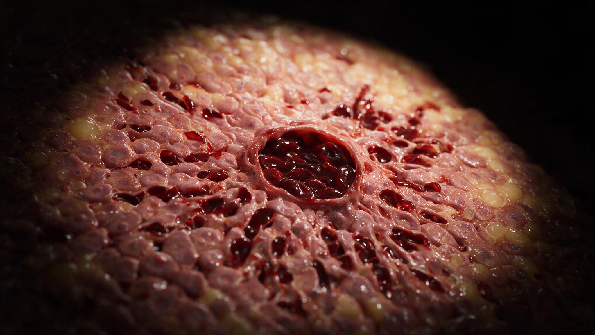
This case study traces the vascular-to-cellular cascade: from the physics of portal pressure overload through the final architecture of coagulative necrosis, where cells retain their ghost outlines even after the cytoplasm has been destroyed. Each panel moves from the macroscopic anatomy of the portal system down to the microscopic landscape of dying hepatocytes.
Portal hypertension begins when resistance to portal blood flow rises. The most common cause is cirrhosis — a stiff, fibrotic liver that acts as a dam. Normal portal pressure runs between 5 and 10 mmHg. When it climbs past 10, the system is hypertensive. When it exceeds 12, complications begin. The portal vein, which normally carries blood from the gut, spleen, and pancreas toward the liver for filtration, now faces a closed door. Blood backs up. Collateral channels that have been closed since fetal life begin to reopen.
The body does not give up. When the liver blocks forward flow, blood finds alternate routes back to the heart. Esophageal varices develop at the gastroesophageal junction. Caput medusae appears around the umbilicus. Hemorrhoids form in the anorectal region. These collaterals relieve pressure, but at a cost: they are thin-walled, fragile, and prone to rupture. More importantly, they bypass the liver entirely, meaning toxins that should have been filtered now circulate freely. But the silent damage happens deeper — at the tissue level, where sustained ischemia from this pressure overload sets the stage for coagulative necrosis.
Coagulative necrosis is the characteristic pattern of cell death following ischemia in most solid organs. Unlike liquefactive necrosis, where enzymes dissolve the tissue into liquid, coagulative necrosis preserves the basic structural outline of the dead cells for days. The cytoplasm becomes eosinophilic and opaque, the nucleus fades or fragments, but the ghost architecture remains. This preservation is important: it means the injury pattern is readable. Under the microscope, the hepatocytes around the central vein show the hallmarks — loss of nuclear detail, preserved cell outlines, an eosinophilic blush where cytoplasm once was. The portal hypertensive liver is one of the best organs to teach this pattern, because the gradient of injury from the portal triad outward is visible.
The teaching goal of this article is twofold. First, to show that portal hypertension is not just a vascular disease — it is a tissue disease. The pressure damages the microvasculature, which starves hepatocytes of oxygen, which triggers the stereotyped pattern of coagulative necrosis. Second, to make coagulative necrosis legible as a pattern: ghost outlines, preserved architecture, eosinophilic cytoplasm, absent nuclei. When a student sees this pattern on a slide, they should think backward through the cascade. What caused the ischemia? What caused the pressure overload? What caused the liver to stiffen in the first place? The illustrations in this series are built to connect those dots — from the portal vein to the dying hepatocyte, from pressure to necrosis, from the macroscopic to the microscopic.
You're set. We'll send the full write-up to your inbox shortly.