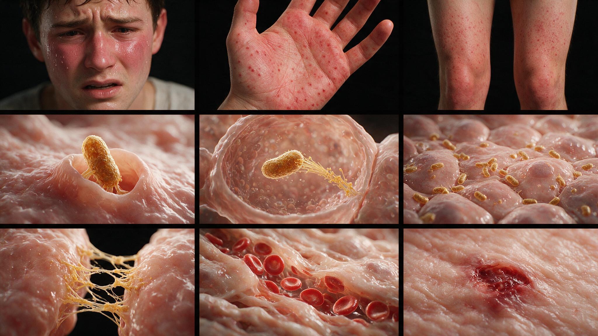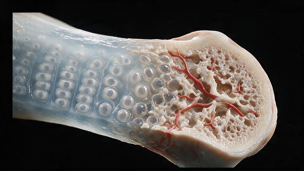
Paget's disease of bone is a disorder of the osteoclast. The cell that normally resorbs bone in a controlled, targeted manner becomes hyperactivated, enlarged, and filled with nuclei — up to 100 nuclei per cell, compared to the normal 3 to 5. These giant osteoclasts resorb bone at an accelerated rate, creating deep resorption pits and triggering a frantic osteoblastic response that lays down new bone just as fast. The result is not stronger bone but disordered bone: architecturally chaotic, mechanically weak, and prone to deformity, fracture, and malignant transformation. The histologic hallmark is the mosaic pattern — irregular, interlocking pieces of lamellar bone separated by prominent cement lines, resembling a jigsaw puzzle assembled by someone who did not have the box for reference. Normal lamellar bone is organized in parallel sheets with uniform cement lines. Pagetic bone is a patchwork of microinfarcts, woven bone, and irregular lamellae, all thrown together at high speed by osteoblasts trying to keep up with osteoclasts that will not stop.
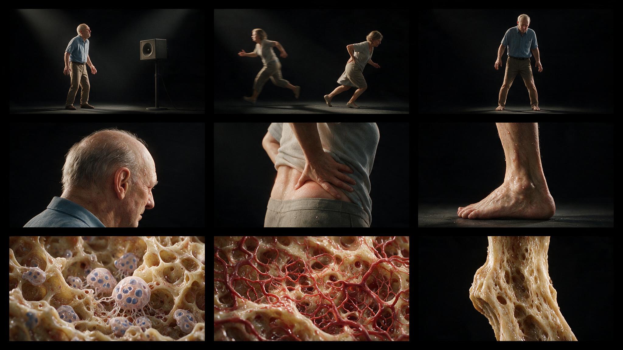
The disease has a striking geographic distribution: it is most common in England, Australia, New Zealand, and western Europe, rare in Scandinavia and Asia. Within a single country, prevalence can vary by region, suggesting an environmental trigger superimposed on genetic susceptibility. The most consistently identified genetic association is with mutations in the SQSTM1 gene, which encodes p62, a scaffold protein involved in RANK-NF-kappa-B signaling in osteoclasts. But the precise trigger that converts a genetically susceptible osteoclast into a pagetic one remains unknown. Some evidence points to paramyxovirus infection (measles, canine distemper virus) of osteoclast precursors as a possible initiating event, but this remains controversial. What is not controversial is the clinical picture: bone pain, deformity (bowing of the tibia, enlargement of the skull), fractures through pagetic bone, and in about 1 percent of cases, the development of osteosarcoma within a pagetic lesion. The diagnosis is made by X-ray (lytic lesions, thickened cortex, coarsened trabeculae) and confirmed by a markedly elevated serum alkaline phosphatase — the enzyme osteoblasts secrete as they furiously lay down new bone.
Paget's disease progresses through three radiologic and histologic phases, though all three can be present simultaneously in different parts of the same bone. The lytic phase (osteitis deformans) is dominated by osteoclast activity: giant multinucleated osteoclasts carve out large resorption lacunae, producing radiolucent regions on X-ray. In the skull, this produces osteoporosis circumscripta — well-defined lytic lesions that look like someone took a hole punch to the calvarium. The mixed phase follows, with both osteoclastic resorption and osteoblastic deposition occurring at high rates side by side. The bone develops a disorganized trabecular pattern and a thickened but porous cortex. The sclerotic phase is the end stage: osteoblast activity outpaces resorption, producing dense, thickened, but mechanically inferior bone. The alkaline phosphatase level mirrors disease activity, rising in the lytic and mixed phases and falling as the disease burns out into the sclerotic phase. A patient with a normal alkaline phosphatase and no bone pain has inactive disease and does not need treatment.
Bisphosphonates are the first-line treatment for Paget's disease, and their mechanism of action explains why. These drugs are pyrophosphate analogs with a P-C-P backbone that resists enzymatic hydrolysis. When administered intravenously (zoledronic acid) or orally (alendronate, risedronate), they bind avidly to hydroxyapatite crystals at sites of active bone resorption. Osteoclasts ingest the bisphosphonate-coated bone matrix during resorption. Once inside the osteoclast, nitrogen-containing bisphosphonates (zoledronic acid, alendronate, risedronate) inhibit farnesyl pyrophosphate synthase in the mevalonate pathway, blocking the prenylation of small GTPases essential for osteoclast function. The osteoclast loses its ruffled border, detaches from the bone surface, and undergoes apoptosis. A single infusion of zoledronic acid (5 mg IV over 15 minutes) normalizes alkaline phosphatase in 80 to 90 percent of patients, with remissions lasting years. The goal of treatment is not to cure the disease — there is no cure — but to suppress bone turnover, reduce pain, prevent progression of deformity, and bring the alkaline phosphatase down to the normal range.
The most feared complication of Paget's disease is sarcomatous transformation — the development of osteosarcoma (or, less commonly, chondrosarcoma or fibrosarcoma) within pagetic bone. The risk is approximately 1 percent over a lifetime for patients with extensive disease, which is hundreds of times higher than the background rate of osteosarcoma. The sarcoma typically presents as a new, rapidly worsening pain in a bone that was previously stable, often accompanied by a soft tissue mass and a lytic lesion on X-ray that looks different from the background pagetic changes. The prognosis is poor: pagetic osteosarcoma is highly aggressive, and five-year survival is less than 10 percent. The mechanism is thought to be the accumulation of DNA damage from the relentless cycle of resorption and formation — each round of remodeling creates opportunities for replication errors, and the hyperproliferative environment selects for clones that have lost tumor suppressor function. Paget's disease is, in this sense, a long-running mutagenesis experiment conducted on the patient's own skeleton.
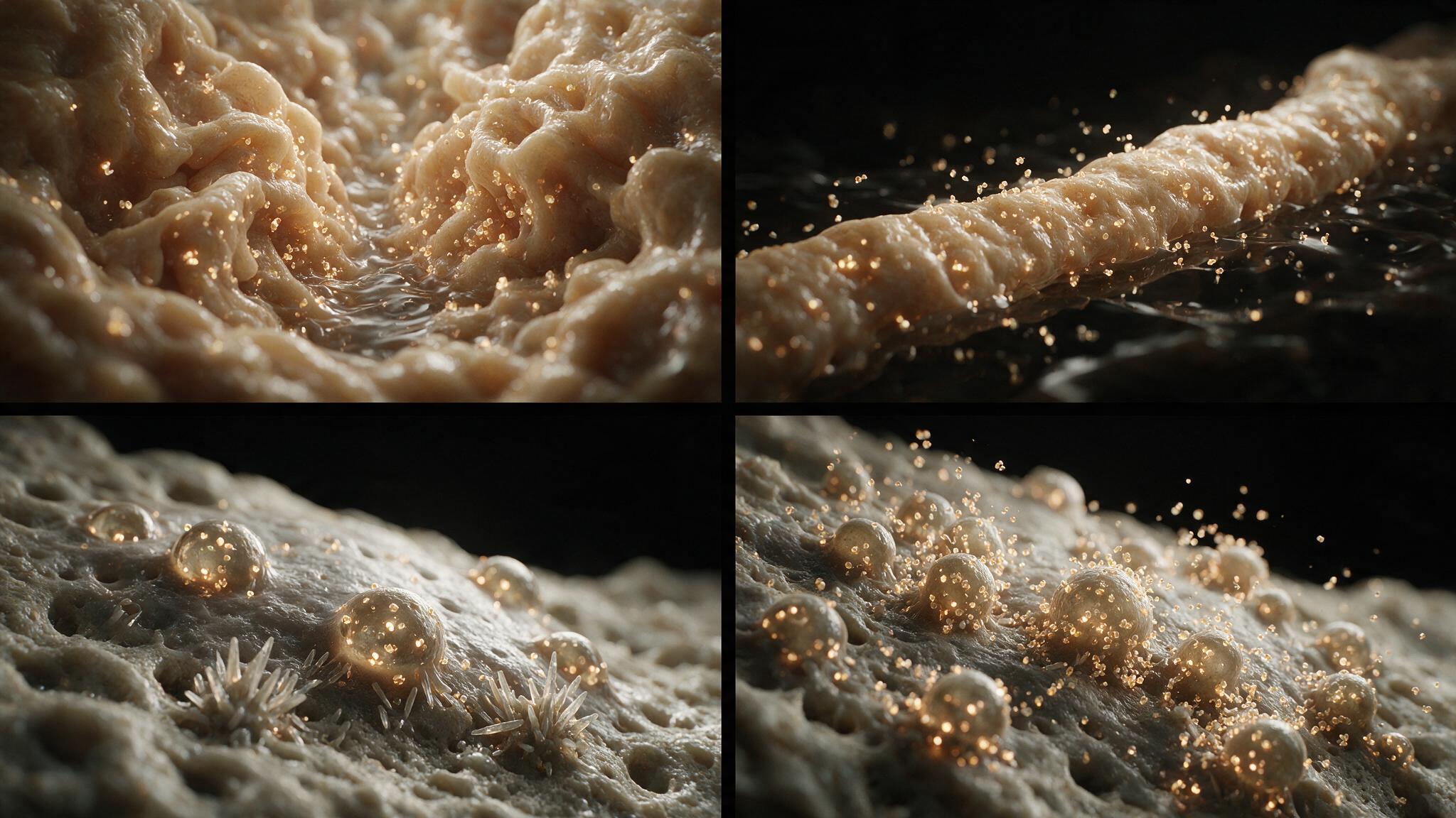
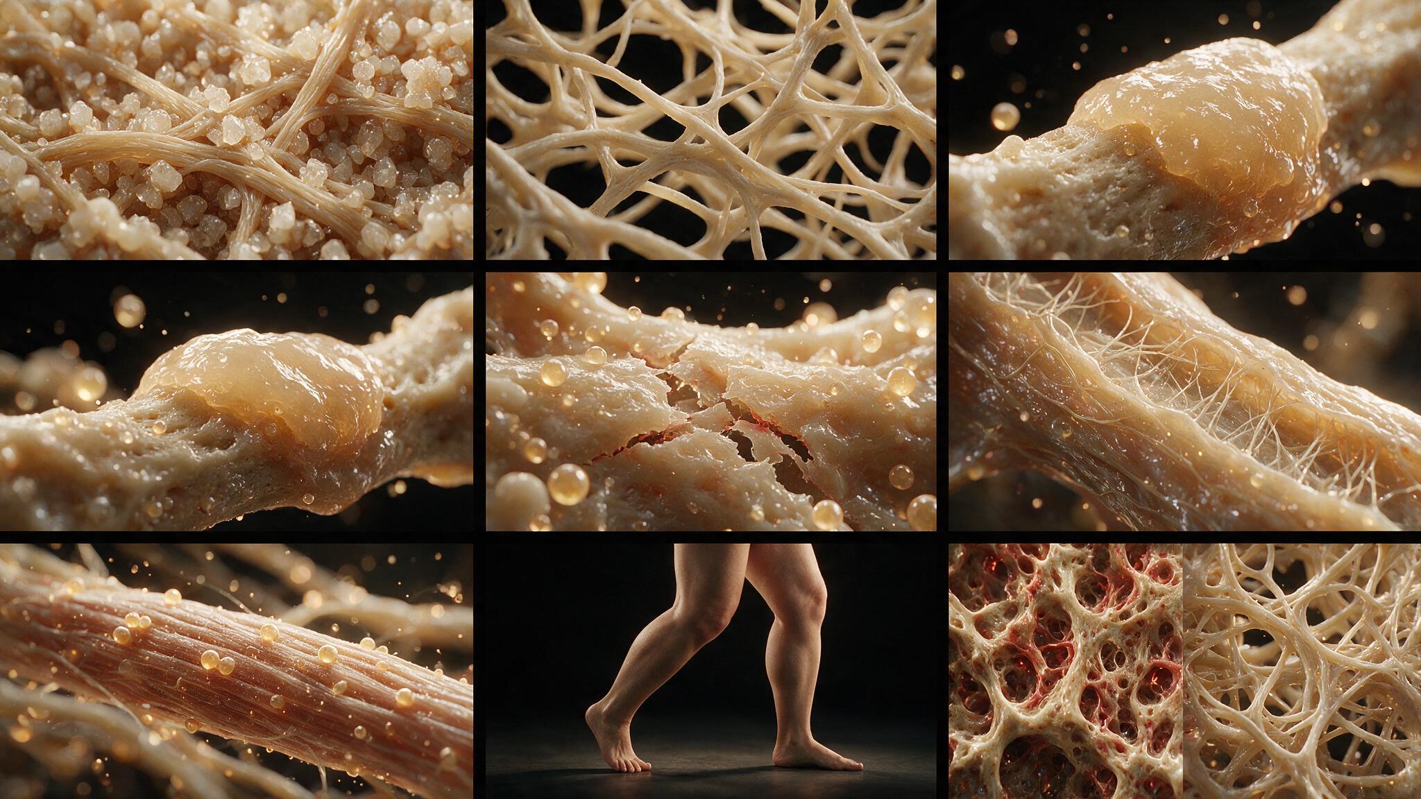
Paget's disease is a disease of bone remodeling run amok. The osteoclast is the primary culprit — hyperactivated, multinucleated, and genetically primed for excessive resorption. The osteoblast follows, laying down new bone as fast as the osteoclast tears it up, but the result is not normal bone. It is disorganized, crisscrossed with cement lines, mechanically inferior, and prone to deformity and malignant transformation. A single infusion of zoledronic acid can suppress the disease for years by poisoning the osteoclast at the moment of resorption, interrupting the cycle and allowing the bone to quiet down. The alkaline phosphatase falls, the bone pain recedes, and the patient's skeleton, for a time, returns to a normal pace of remodeling. But the risk of sarcoma never goes to zero, and the deformities already acquired do not reverse. This illustration series renders Paget's disease from the gene pool to the domestic scene: the molecular drivers in the osteoclast nucleus, the chaotic mosaic of the histology section, the bowed tibia and enlarged skull in the clinic, and the rebar-like disorganization that makes pagetic bone a poor structural material.
You're set. We'll send the full write-up to your inbox shortly.