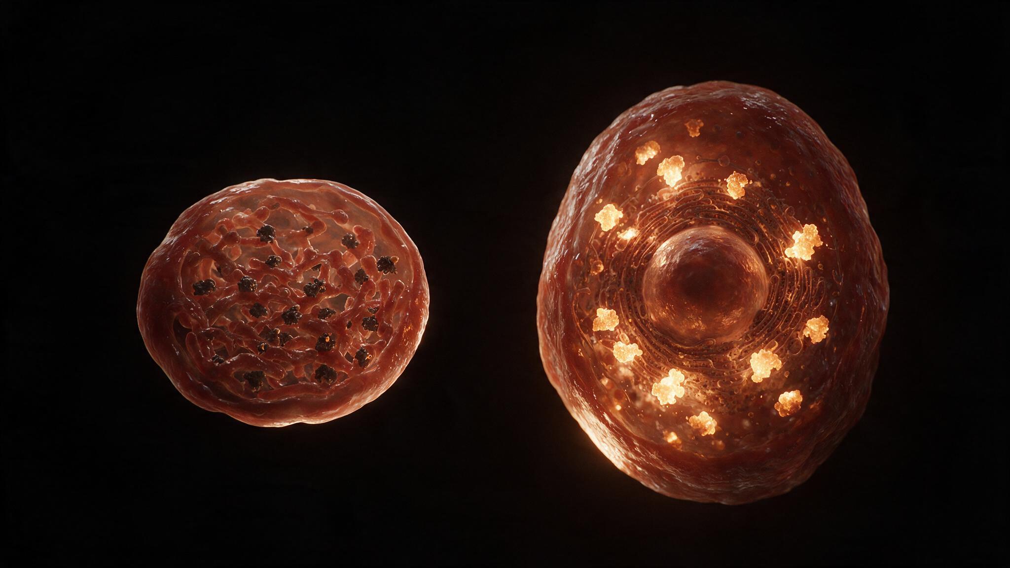

The neuron at rest is a battery: -70 mV across a membrane only 5 nanometers thick, maintained by the sodium-potassium ATPase pumping three sodium ions out for every two potassium ions in. This gradient is potential energy stored in ion concentration differences across a lipid bilayer that is, at rest, essentially impermeable to sodium. When a stimulus depolarizes the membrane to threshold — roughly -55 mV — voltage-gated sodium channels in that patch of membrane detect the change. Their S4 voltage-sensing helices, studded with positively charged arginine residues, are physically pulled outward by the changing electric field. The channel's central pore opens. Sodium floods inward down its electrochemical gradient. The membrane potential reverses from -70 mV to +30 mV in under a millisecond.
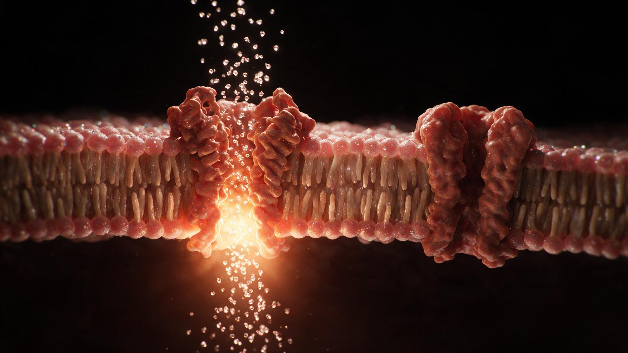
This is the action potential: a self-amplifying, all-or-none electrical impulse that propagates without decrement along the axon at speeds up to 100 meters per second. Local anesthetics like lidocaine bind to the intracellular mouth of this same sodium channel and block the pore. Tetrodotoxin, from pufferfish, binds the extracellular mouth. Anti-epileptics like phenytoin stabilize the inactivated state. Each of these drugs works because the sodium channel is the single point of failure for neuronal excitation. This case study renders the channel at the molecular level: the open pore, the ion stream, and the charged plasma environment that makes it all possible.
The voltage-gated sodium channel is a single polypeptide of roughly 2,000 amino acids folded into four homologous domains (I-IV), each containing six transmembrane segments (S1-S6). The S4 segment of each domain is the voltage sensor: every third residue is a positively charged arginine or lysine, creating a helical stripe of positive charge embedded in the membrane electric field. At rest, the inside of the cell is negative relative to the outside, so the S4 helices are pulled inward. When the membrane depolarizes — when the inside becomes less negative — the electric force holding S4 inward weakens. The helices slide outward through the protein's gating pore, a movement of only a few angstroms that is mechanically coupled to the S6 segments lining the central pore. As S4 moves out, S6 helices splay apart. The pore opens. This is activation: voltage sensing converted to pore gating in microseconds.
The open pore of the sodium channel is exquisitely selective. It passes sodium ions at near-diffusion-limited rates — roughly 10 million ions per second — while rejecting potassium ions, which have a larger ionic radius. The selectivity filter, a ring of glutamate residues at the narrowest point of the pore, strips water molecules from the sodium ion and coordinates it with precisely spaced oxygen atoms. A potassium ion, larger even when dehydrated, cannot fit through this ring. The driving force for sodium entry is twofold: the concentration gradient (140 mM outside, 10 mM inside) and the electrical gradient (negative inside attracts positive sodium). As sodium floods in, the membrane potential rises from -70 mV toward the sodium equilibrium potential of roughly +60 mV. The rapid influx of positive charge itself depolarizes adjacent patches of membrane, triggering their sodium channels to open. This is the self-amplifying nature of the action potential: sodium entry in one patch depolarizes the next patch, which opens more channels, which depolarizes the next patch. The impulse propagates.
The sodium channel does not stay open. Within a millisecond of opening, the intracellular loop connecting domains III and IV swings into the pore's inner mouth like a ball on a chain, physically occluding it. This is fast inactivation — an auto-shutoff that terminates the sodium current and ensures the action potential is brief and unidirectional. The channel then enters a refractory state from which it cannot reopen until the membrane repolarizes and the inactivation gate resets. Meanwhile, voltage-gated potassium channels, slower to activate, open and allow potassium to flow outward, restoring the negative membrane potential. The sodium-potassium ATPase then slowly pumps the ions back to their resting distributions. The entire cycle — depolarization, sodium channel opening, inactivation, potassium efflux, repolarization — takes about 2 to 3 milliseconds. The channel is then ready to fire again.

The voltage-gated sodium channel is one of the most targeted proteins in pharmacology. Local anesthetics bind to a site in the inner pore and stabilize the inactivated state, preventing the channel from reopening. Antiarrhythmics like flecainide slow recovery from inactivation, reducing the maximum firing rate of cardiac myocytes. Anticonvulsants like phenytoin and carbamazepine preferentially block channels that are firing at high frequency, selectively suppressing seizure activity while leaving normal firing relatively intact. TTX, the pufferfish toxin, binds with picomolar affinity to the extracellular mouth of the channel and blocks sodium entry outright — a single milligram is lethal. Each of these drugs tells the same story from a different angle: the sodium channel is the gatekeeper of excitability, and controlling that gate is the most direct way to control the nervous system, the heart, and the muscles. The illustrations in this series render the channel at the scale where voltage becomes motion, where an electric field physically moves a helix, and where that movement opens a path for ions to change the voltage of a cell.
You're set. We'll send the full write-up to your inbox shortly.